


MRI Takes the Sting Out of Painful Screening Tests: Brain Scans Alone Can Distinguish Between Dementia Types
MRI scans can be used to accurately distinguish different types of dementia without the use of invasive screening tests like a lumbar puncture, a new study suggests.
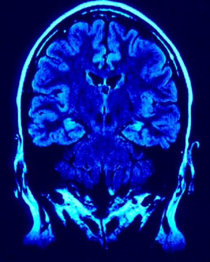
A new study, published Wednesday in the journal Neurology, reveals that structural brain patterns on medical scans could accurately identify Alzheimer's disease and another type of dementia, Frontotemporal Lobar Degeneration (FTLD).
Researchers said that because Alzheimer's disease and frontotemporal lobar degeneration (FTLD) often have similar symptoms, doctors often struggle to diagnose the dementia, which means that treatment may be delayed.
While more invasive tests like cerebrospinal fluid analyses remains the most accurate method for predicting the type of dementia and the underlying cause for the disease, it requires invasive lumbar puncture, which can be unpleasant for patients.
Even though Alzheimer's disease and frontotemporal lobar degeneration (FTLD) are two distinct diseases with very different underlying processes, they share similar clinical features and symptoms that can be hard to distinguish between without tests.
Both the diseases cause the patient to be confused and forgetful and can affect a person's personality, emotions and behavior.
However, Alzheimer's disease generally affects the cerebral cortex, the brain's outer grey matter, and FTLD affects the temporal and frontal lobes of the brain, which can show up on brain scans.
A cerebrospinal fluid test, which requires lumbar puncture, or a needle to be stuck into the spine, can also differentiate between these two diseases by checking protein levels in the brain. Protein levels tend to be higher in patients with Alzheimer's disease than in those with FTLD.
Because lumbar puncture can be painful for patients, researchers from University of Pennsylvania wanted to see if they can eliminate the agonizing needle-to-spine altogether and just use MRI brain scans to predict brain protein levels.
Researchers examined the brain scans of 185 patients who had already been diagnosed with Alzheimer's disease or FTLD and had undergone a lumbar puncture test as well as a MRI scanning to see if they could identify any patterns that corresponded with protein level results from lumbar puncture tests.
The findings reveal that the density of gray matter on the MRI scans correlated with protein results and that the new MRI test developed by researchers identified the correct diagnosis 75 percent of the time.
Researchers said that the results show that the new MRI prediction method had a similar accuracy rate as the lumbar puncture method, and while the accuracy rate is not 100 percent, the new MRI prediction method could still be a useful screening tool.
"This could be used as a screening method and any borderline cases could follow up with the lumbar puncture or PET scan," Lead researcher Dr. Corey McMillan said in a statement.
"This method would also be helpful in clinical trials where it may be important to monitor these biomarkers repeatedly over time to determine whether a treatment was working, and it would be much less invasive than repeated lumbar punctures," he concluded.


