


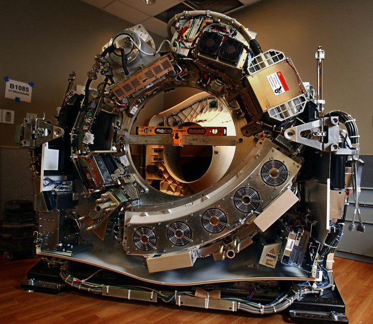
CT Scanner Without Its Cover Demonstrates How It Works; Inside The Machine That Reveals Your Insides
Doctors can look inside a patient’s body by requesting scans of bones, blood vessels, brains, and soft tissue thanks to the computer tomography (CT) scanner. After the first clinical CT scan of a patient was performed in 1971, radiologists have used scans to diagnose tumors, traumas, and plan medical, surgical, and radiation treatment on nearly all parts of the human body.
Inside the doughnut-shaped machine, where patients lie on the table as it slowly moves through the scanner, the machine spins around. As it spins, it sends a thin X-ray beam through the body, which is collected on the other side of the machine and transferred to computer software, where it is uploaded on a screen for CT technologists to view. It converts into a cross-sectional picture, scan, or “slice” of the human body, taking at some sessions fewer than 15 minutes to complete the CT scan. It’s become a reliable and routine medical practice over the years. From 1996 to 2010, CT scan rates have tripled, adding up to 149 scans per every 1,000 patients who enter a hospital.


