


Stem Cells' 'Black Box' Captured For The First Time, Explaining Once-Mysterious Process
Using time-lapse imaging and stem cells dyed green, a team of scientists from Boston Children's Hospital's Stem Cell Research Program has uncovered several clues that may advance the treatment of bone marrow cancer, severe immune deficiencies, and blood disorders.
For roughly a decade, science has known the stem cells found in bone marrow can be transplanted from donor to patient, but what wasn’t clear was how that transition happened. In a new study to be published in the journal Cell, the team details its findings on zebrafish, which provide a reliable model for understanding how human stem cells work. With the newfound access, scientists are confident they can launch more specific treatments in the fight against certain cancers.
"The same process occurs during a bone marrow transplant as occurs in the body naturally," said Dr. Leonard Zon, senior investigator and director of the Stem Cell Research Program, in a statement. "Our direct visualization gives us a series of steps to target, and in theory we can look for drugs that affect every step of that process."
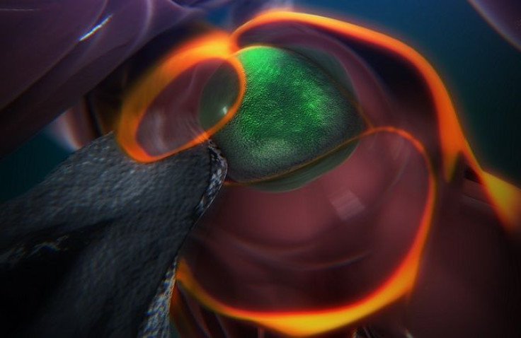
Blood stem cells live in bone marrow and are ultimately responsible for creating blood. The challenge for researchers is that the cells can only perform that function once they’ve found a "niche." How that niche forms is a point of discovery for Zon and his team, and with the help of standout green dye they can see the niche developing in the zebrafish’s tail. The cells attach to the blood vessel wall, before a chemical signal instructs it to push through to the other side. Then the magic happens.
"In that space, a lot of cells begin to interact with it," Zon said. Specifically, blood-vessel cells known as endothelial cells begin to swarm the stem cell. "We think that is the beginning of making a stem cell happy in its niche, like a mother cuddling a baby." Soon after, a separate "nurse" cell, otherwise known as a stromal cell, helps the stem cell stay attached, until it eventually divides and begins the process over again to populate the desired location in the body.
Zon and his team have assembled the images into an animation that details the inner-workings of the process. "Nobody's ever visualized live how a stem cell interacts with its niche," he said. In follow-up tests on mice, the team found similar outcomes. This suggested mammals and fish, and perhaps even humans, depend on these pathways. Ultimately, they will use the information to develop more advanced ways to carry out bone marrow transplants.
"Stem cell and bone marrow transplants are still very much a black box — cells are introduced into a patient and later on we can measure recovery of their blood system, but what happens in between can't be seen," said Dr. Owen Tamplin, the paper's co-first author. "Now we have a system where we can actually watch that middle step."
Source: Durand E, Carr L, Hagedorn E, Lee P, Zon L. Cell. 2015.


