


3D Brain Model Of Most Studied Amnesiac, Henry Molaison, Provides Insight Into Human Memory
Every neuroscience textbook mentions one very famous patient who was identified by his initials, H.M., up until his death in 2008. Recently, the brain of the most studied amnesic patient in the history of neuroscience, Henry Molaison, has undergone a detailed postmortem analysis that will provide histological and digital 3D insight into a case that profoundly shaped our understanding of human memory.
Molaison blew open the doors of modern neuroscience in 1953 when he had brain surgery at the age of 27 to reduce severe epileptic fits. No one at the time knew that surgically removing certain regions of his brain would result in an inability to remember daily events or recognize hospital staff that took care of him throughout the day. Doctors were perplexed that his personality, language ability, and perceptual skills remained unchanged despite an inability to store information in his long-term memory. His IQ even remained above average 10 months after the operation.
Neuropsychologists, such as Brenda Milner of McGill University, came to understand that Molaison’s near-total amnesia stemmed from removing a region of the brain that’s now understood to be crucial for memory formation, the medial temporal lobe. “The purity and severity of H.M.’s memory impairment, combined with his willingness to participate in testing, made his case study uniquely influential in the field of memory research,” explained University of California San Diego neuroanatomist, Jacapo Annese, and colleagues in a study published in Nature Communications.
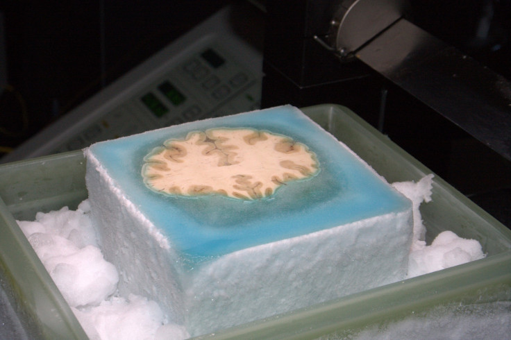
In the journal, the researchers report the results of dissecting Molaison’s brain into 2,401 thinner-than-paper slices that were each captured as a digital image before being cryogenically preserved in serial order. The images have been archived and used to create a 3D model of the entire brain, which provides an unprecedentedly detailed atlas of Molaison’s brain and the surgery that affected his memory the way it did. “Our goal was to create a detailed three-dimensional … model of the whole brain from high-resolution anatomical images so that we could revisit, by virtual dissection, [the] surgical procedure and localize the anatomical borders of the lesion in the temporal lobe,” Annese and colleagues explained.
Indeed, this detailed analysis revealed for the first time that part of the so-called orbitofrontal cortex, which is a brain region that’s involved with decision-making, had a small lesion that Annese suspects was inflicted when the original surgery occurred. This insight might be one of many to come thanks to archived series of brain images being of a much higher-resolution than the non-invasive MRI scans that were previously conducted on Molaison. Perhaps, the archive might further explain the nuances of his inability to remember having an exchange with a person he had a proficient conversation with minutes earlier yet still be able to learn a new motor task.
Overall, the authors concluded, “The archive of images will … constitute a permanent digital trace for the case that defined two generations of memory research and that will likely remain as emblematic for neuroscience in the future as it is today.”
Source: Annese J, Schenker-Ahmed N M, Bartsch H, et al. Postmortem examination of patient H.M.’s brain based on histological sectioning and difital 3D reconstruction. Nat Commun. 2014.
Published by Medicaldaily.com


