Autism Doesn’t Change Brain’s Anatomy Most Of The Time, Only Its Function

Going against hundreds of studies that argue in defense of real, anatomical differences between people with autism spectrum disorder (ASD) and people without it, a new large-scale study suggests many modest differences aren’t compelling enough to take seriously.
The findings imply a far deeper set of truths to ASD than prior research has led on: People with ill-functioning brains don’t exhibit a different set of temperaments and behaviors because of any broad, surface-level discrepancies on an anatomical level, but rather a finely tuned set of functional, or neuropathological, differences. If upheld in future research, the findings could close a major door for ASD.
“Substantial controversy exists regarding the presence and significance of anatomical abnormalities in autism spectrum disorders,” the researchers wrote in their report. Indeed, while MRI studies (along with the present study) have found slight differences in the structural anatomy of certain ASD subjects, much of the research is highly fragmented and not easily translated to other investigations.
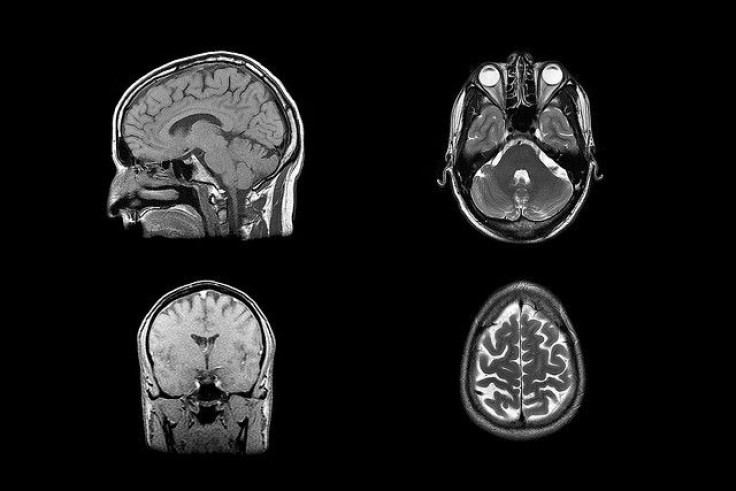
In their current study, a group of Israeli researchers collected data from 539 people diagnosed with high-functioning ASD and 573 controls — relying on the samples collected from the publicly available Autism Brain Imaging Data Exchange. Comparing the samples, the team saw no differences in brain region size among the two groups nor any differences in total brain volume. The only significant increase was in ventricle size, though many disorders in addition to ASD have been found to cause this effect.
More relevant to the discussion of brain anatomy and ASD is the thickness of the brain’s cortex. Some research has found that cortical thickness — in other words, how thick the folds are — may determine the degree to which a person suffers social impairments. In the current study, these findings were minimal:
Individuals with ASD exhibited significantly thicker cortex than controls in several areas, including the right and left occipital poles, left middle occipital sulcus, left occipital-temporal sulcus… [however] there were no significant differences in volumetric or surface area measures between groups. There were also no significant volumetric differences between groups in any of the subcortical areas.
But observational analysis via computer software wasn’t enough for the researchers. They wanted to try their hand at telling the brains apart themselves. So they turned to a machine learning technique called linear discriminant analysis, which lets scientists pick apart the individual differences on their own. Even here, the best results fell at a paltry 60 percent. Other methods of analysis yielded a 50 percent accuracy score.
The upshot? The team found no differences. And their own methods amounted to little more than scientific coin-flipping.
“This suggests that anatomical measures alone are likely to be of low scientific and clinical significance for identifying children, adolescents, and adults with ASD or for elucidating their neuropathology,” the team concluded. But being able to make these identifications is of course important, particularly as one in 68 children in the U.S. is currently diagnosed with autism.
If the findings can be replicated on a similarly large scale — especially important since the impact diminished when the researchers purposely shrank their sample size — then the scientific community as a whole may benefit from newfound understanding. At least for now, the data seem to suggest we’re looking in the right direction, just not as closely as we should be.
Source: Haar S, Berman S, Behrmann M, Dinstein I. Anatomical Abnormalities in Autism? Cerebral Cortex. 2014.


