


Bacterial Pneumonia May Lead To A Heart Attack When Pathogens Invade And Kill Muscle Cells
Pneumonia is often described as “the old man's friend” because it can provide a peaceful and dignified death to someone who is elderly and suffering from other more serious conditions, such as cancer. However, there is another face to pneumonia, which is cruel and quite literally heartless: in adults of all ages, bacterial pneumonia elevates the risk for cardiac trouble, including heart attacks. Now, a new study from the University of Texas demonstrates exactly how Streptococcus pneumoniae, responsible for most cases of bacterial pneumonia, invades the heart and causes the death of heart muscle cells. Having gained a better understanding of the interaction between pathogen and our hearts, scientists may work toward better treatments and prevention strategies to protect the heart from damage incurred during this illness which affects millions of people each year.
Pneumonia is an inflammation of the lungs usually caused by infection with bacteria, viruses, and other organisms. Simple triggers, such a case of the flu or a simple respiratory infection, may lead to pneumonia when a patient's defense system is weakened. With antibiotic treatment (except for cases of viral pneumonia), most patients improve within a couple of weeks.
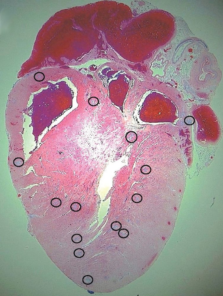
However, in some cases, patients develop heart problems and such “cardiac complications were implicated as a direct or underlying cause of death in 27 percent of the pneumonia-associated deaths,” wrote the researchers of the current study. To better understand how pneumonia may result in heart problems, Dr. Carlos Orihuela of the Health Science Center at UT San Antonio and his colleagues began their research into invasive pneumococcal disease by focusing on mice. Examining the infected mice, the scientists observed elevated levels of troponin, a marker for heart injury, in their blood as well as abnormal EKGs. Further investigation of their hearts showed microscopic sites of injury (or microlesions) in the muscle where S. pneumoniae were found, proving the bacteria had invaded and multiplied there. In fact, the researchers identified dying heart muscle cells in the tissue surrounding the microlesions.
What, then, had occurred at the molecular level? The S. pneumoniae toxin pneumolysin, found within the microlesions, was responsible for heart muscle cell death, and, the researchers discovered, had traveled there with the help of a protein called CbpA (choline-binding protein A). In part, CbpA creates its adverse effects by inducing the release of pro-inflammatory molecules.
To further investigate this process, the researchers obtained tissues from both rhesus macaques and humans who had died from pneumococcal pneumonia. In both the monkeys and humans, the team observed cardiac microlesions similar in size and appearance to those seen in mice, but without the presence of S. pneumoniae bacteria. However, a key difference existed: the macaques and the human patients had been treated with antibiotics, while the mice had not. Could the bacteria have caused the lesions but subsequently been killed by the treatment?
To test this theory, the researchers infected mice with S. pneumoniae and treated them with Ampicillin, a high-dose antibiotic, when the lesions first became apparent. This time the hearts of the infected mice, like those of the macaques and humans, showed microlesions devoid of bacteria. Having demonstrated how S. pneumoniae can compromise the heart, the researchers suggest alternative antibiotics to Ampicillin, which breaks bacteria apart and causes them to release their contents (including pneumolysin), would be more advantageous when treating patients with pneumonia.
Source: Brown AO, Mann B, Gao G, et al. Streptococcus pneumonia Translocates into the Myocardium and Forms Unique Microlesions That Disrupt Cardiac Function. PLOS ONE. 2014.
Published by Medicaldaily.com


