


Breast Cancer Imaging Tool Detects Fine Lines Between Healthy And Diseased Tissue
Cancer researchers from Brigham and Women’s Hospital (BWH) have developed and tested a tool for finding cancerous tissue, which they believe could reduce the need for follow-up tissue removal surgeries by helping doctors remove the diseased tissue all at once.
Published in the Proceedings of the National Academy of Sciences, the new study utilizes a diagnostic tool known as Desorption ElectroSpray Ionization mass spectrometry imaging, or DESI, for short. While mass spectrometry imaging has been in use for years, the new technique relies on the novel conversion of molecules into ions — a process known as ionization — to detect cancerous tissue.
“Our findings demonstrate the feasibility of classifying cancerous and normal breast tissues using DESI mass spectrometry imaging,” said Dr. Nathalie Agar, director of the Surgical Molecular Imaging Laboratory and the study’s senior author, in a statement.
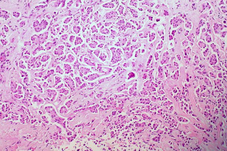
Tests using traditional pathology confirmed the technique’s accuracy. When Agar and her colleagues at BWH looked at the distribution of lipids within breast tissue and normal tissue from 61 samples of 14 breast cancer patients, post-mastectomy, they found several fatty acids were more present in the tissues with cancer. A software program helped demarcate the boundaries between diseased and healthy tissue.
In future research, Agar and her team look to enhance the detection and validation of these lipids as biomarkers for cancer. In the meantime, the best science can do for diagnosis is to take an X-ray of the tissue through use of mammography, and take invasive follow-up biopsies to check for disease under the microscope. For as far as the methods have advanced, they are still imperfect. And when surgeons don’t know how much tissue to remove, they may leave some behind.
“The results may help us to move forward in improving this method so that surgeons can use it to rapidly detect residual cancer tissue during breast cancer surgery,” Agar said, “hopefully decreasing the need for multiple operations.”
In the U.S. alone, one in eight women will develop invasive breast cancer over the course of her lifetime. Fortunately for the some 230,000 new cases that arose in 2014, the death rate from breast cancer has been falling steadily since 1989, according to data from the Centers for Disease Control and Prevention. Experts suspect the reductions stem from increased awareness, treatment advances, and earlier screening.
The research team also hopes to bring DESI to its Advanced Multimodality Image Guided Operating suite, which would help surgeons determine breast cancer’s margins more effectively. In this sense, “margins” refers to the distance between healthy tissue and tissue that’s been overrun with cancer cells. If the team can hone its technique to the point it can make real-time assessments, surgeons would no longer need to make second incisions — a win for an already invasive form of treatment.
Source: Calligaris D, Caragacianu D, Liu X, et al. Application of desorption electrospray ionization mass spectrometry imaging in breast cancer margin analysis. Proceedings of the National Academy of Sciences. 2014.


