


‘Cancer Goggles’ Help Surgeons See Malignant Tumor Cells; New Technology Will Be Tested In Dogs
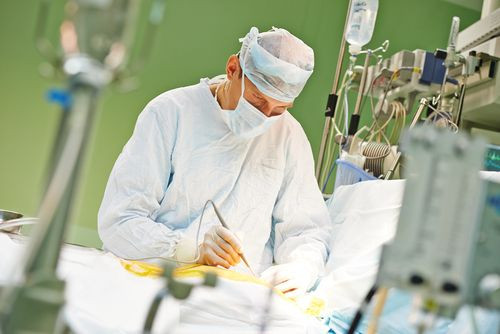
Cancer cells are difficult to see, which makes each surgery to remove a tumor — and every last cancer cell — especially difficult and particularly fraught. Dr. Samuel Achilefu, director of the Optical Radiology Lab and a professor of biomedical engineering at Washington University, is in the process of developing "cancer goggles" that would help a surgical team see malignant tumor cells during an operation. Although the goggles are still in the development stage, many believe this new technology, which is supported by grants from both the National Cancer Institute and the National Institute of Biomedical Imaging and Bioengineering, will be a boon for surgeons and patients alike. After all, if the use of these goggles is able to reduce follow-up surgeries, they would also eliminate a great deal of anxiety, suffering, and pain on the part of patients.
How did the new technology come about? As Achilefu told Columbia Daily Tribune, a former surgical fellow asked him if it was possible to create a device that would help surgeons accurately visualize human tissue while still in the operating room. After all, CT scans and MRIs provide doctors with terrific maps that allow them to see where the cancerous tumors are located. Yet once they enter the operating room and cut into the tissues of an actual patient in real-time, the usefulness of those static images fades. A surgical team, the fellow explained, finds it extremely difficult to distinguish cancer cells from healthy cells during an actual operation, and for this reason, they find it hard to ensure they’ve removed all the malignant tissue. This is why surgeons often remove extra tissue as a margin of error and send these samples to a pathology lab to check for the presence of cancer. Unfortunately, when cancer cells are detected, a second surgery is recommended.
Using the new technology, doctors will be able to "see" the malignant tissue with the help of a contrasting agent, Biomarker LS 301, injected into a patient before the surgical procedure. LS 301 enters cancer cells, staining the infected tissue from the inside out and creating a bright fluorescent glow when an infrared light, shining from a small monocle on the goggles, hits them. Oddly, LS 301 was a mistake, Achilefu told The Beacon, as he was trying to create the exact opposite — a control agent that would not go into cancer cells. Eventually, understanding it could selectively bind to breast, prostate, liver, brain, colon, leukemia, and lymphoid cancers, and even stayed in these malignant cells, including stem cells, for about a week, Achilefu thought LS 301 might be useful for some other purpose.
Achilefu, who is head researcher of the cancer goggles project, said he hopes his new goggles will help surgeons differentiate between normal and cancerous tissue and so enable them to remove it all at once. Yet before the goggles can be used or even tested on humans, the goggles will be tested by researchers at the Missouri University veterinary school on dogs. Jeffrey Bryan, associate professor of oncology in veterinary medicine and surgery, told The Maneater the school often works with breast cancer in dogs. Similar to humans, dogs often develop both benign and malignant tumors yet even more importantly, tumors develop and behave very similarly to those in humans. The clinical tests in dogs are slated to begin shortly.
Published by Medicaldaily.com


