


Cancer Research Gets An Upgrade; 3D Microscope Allows Us To See Diseases Like Never Before
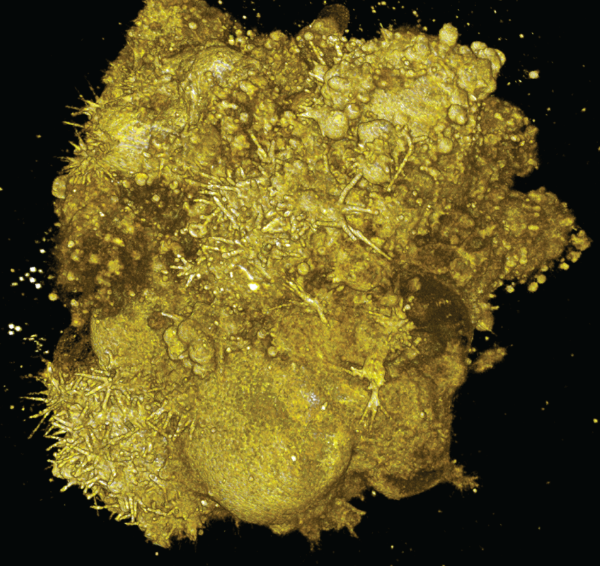
Cancer cells in the body often behave differently than when they’re beneath the glass of a microscope slide. To study them, however, it’s crucial that scientists see them in their natural environment — something that’s been difficult to achieve. In a recent study, however, a team of United States scientists built a high-resolution microscope that captures 3D images of cancer cells in hopes that it will give us a better understanding of how the cells act in our bodies.
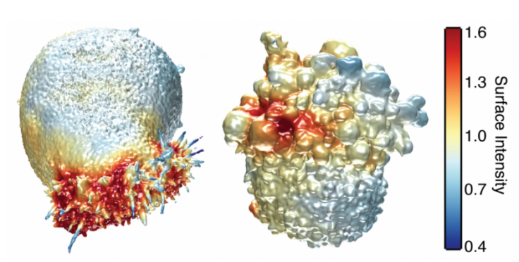
It’s well known to researchers that cancer cells move and interact with one another differently when tamped down under a slide. Effective 3D microscopes, however, have been hard to develop; they must be precise enough to capture a detailed image without interfering with the cells’ microenvironments. Likewise, scientists should be able to manipulate the microenvironments without using chemical or mechanical influences. A team of researchers from the University of Texas Southwestern Medical Center devised a solution with their high-resolution microscope, which can simultaneously image the cells and the collagen matrix they're embedded in.
“There is clear evidence that the environment strongly affects cellular behavior — thus, the value of cell culture experiments on glass must at least be questioned," said senior author Reto Fiolka, an optical scientist at the UT Southwestern Medical Center, in a statement. "Our microscope is one tool that may bring us a deeper understanding of the molecular mechanisms that drive cancer cell behavior, since it enables high-resolution imaging in more realistic tumor environments."
One particular way in which the 3D microscope might help scientists is by exposing the purpose of small protrusions on the surface of tumors, called blebs. The researchers hypothesize that these blebs play a role in helping cancer cells survive or move around. By studying them in skin cancer cells, the researchers might gain a better understanding of why some people never develop skin cancer, or why some patients with the disease develop resistance to treatments.
Now that the team has designed and created the microscope, co-first author Erik Welf says it’s time to work toward making “meaningful contributions to cancer cell biology.” However, because the images the microscope produces are still too complex to interpret with the human eye, the team will first have to design a software that’s advanced enough to analyze the images.
Source: Welk ES, Driscoll MK, Dean KM, et al. Quantitative Multiscale Cell Imaging in Controlled 3D Microenvironments. Developmental Cell . 2016
Published by Medicaldaily.com


