Developments In Bone Grafting: New Synthetic Graft Will Reduce The Number Of Additional Surgeries
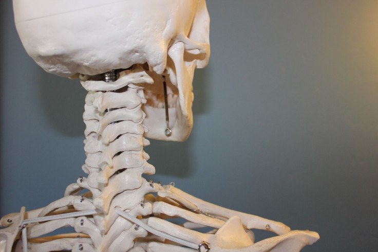
A new type of bone graft is on the horizon, thanks to researchers led by the Queen Mary University of London (QMUL). The scientists have developed a new type of synthetic bone that enhances the body’s own ability to regenerate bone tissue, and it could improve outcomes for patients.
The study detailing the research, published in the Journal of Materials Science: Materials in Medicine, explained that the new graft — called Inductigraft — was able to guide bone tissue regeneration in as little as four weeks. Defects were evaluated at four, eight, and 12 weeks, and the new graft’s performance matched up with that of the clinical gold standard, called autograft. Autograft is made up of the patient’s own bone, containing living cells and growth factors.
Bone grafts are a surgical procedure used to fix bones damaged from trauma, or help with problem joints. It is also useful for helping bone grow around implanted devices, such as a knee replacement.
“Our challenge is to develop a graft that’s as clever as bone,” said Dr. Karin Hing, co-author of the study and reader in Biomedical Materials at QMUL’s Institute of Bioengineering, in a statement. “For this synthetic graft, we looked at the mechanics of how bone adapts to its environment and changed both the chemical and physical composition of the graft, specifically how the holes within the structure are placed and interconnected.”
The researchers manipulated the pore structure of the graft to best imitate natural bone tissue. Hing said that the study has major implications for those suffering from skeletal injuries, and for surgeons.
“At the moment, the preference is to use the patient’s own tissue to create or enhance bone grafts. However, our results show that Inductigraft can be just as effective, with the advantage that the patient doesn’t have to undergo additional surgery to harvest the autograft.”
This breakthrough builds upon past research conducted at QMUL, where researchers added silicate into hydroxyapatite, a traditional synthetic substitute material containing calcium and phosphate, which is chemically similar to natural bone mineral. This enhanced the graft chemistry, and scientists found that the combination of optimized chemistry and pore structure was better at guiding cells to differentiate into cells that produce bone tissue, in both laboratory and body settings.
More research, funded by two spin-outs from QMUL, is currently underway to examine the mechanism by which Inductigraft is capable of guiding bone formation.
Source: Hutchens S, Campion C, Assad M, Chagnon M, Hing K. Efficacy of silicate-substituted calcium p0hosphate with enhanced strut porosity as a standalone graft substitute and autograft extender in an ovine distal femoral critical defect model. Journal of Materials Science: Materials in Medicine. 2015.


