


Exploring The Brain: New Technology Produces Images Of Neural Connections At Nanoscale Resolution
A new imaging tool allows scientists to generate images of the brain and its many neuronal connections at a previously unattainable nanoscale resolution. While the inventors’ immediate aim is to turn electron microscopy images of the brain into a minable dataset, over the long-term they hope to make the resource available to the scientific community in the form of a national brain observatory. Undoubtedly, this research will be instrumental in efforts to create a detailed map of the human brain.
What is a connectome?
The suffix "-ome" refers to a totality. The human genome, for instance, is the complete set of DNA carried by a single cell in the body. Similarly, the human connectome refers to a complete map of the structural and functional connectivity of the brain. Because neurons in our brains make connections in direct response to our experiences, the new tool is expected to help in the effort to map the connectome, ultimately revealing the similarities and differences among our individual brains. Dr. Narayanan Kasthuri, first author of the study and assistant professor of anatomy and neurobiology at Boston University School of Medicine, speculated the tool might answer a host of questions about individual differences among human brains.
"What parts of brains circuitry are stereotyped and which parts are variable across humans? How do human brains grow up given the dramatic differences in behavior between children and adults?" Kasthuri speculated in an email to Medical Daily. The technology might also help explain neurological disorders by allowing scientists to compare the neuron-to-neuron connections in schizophrenia patients with those in healthy people.
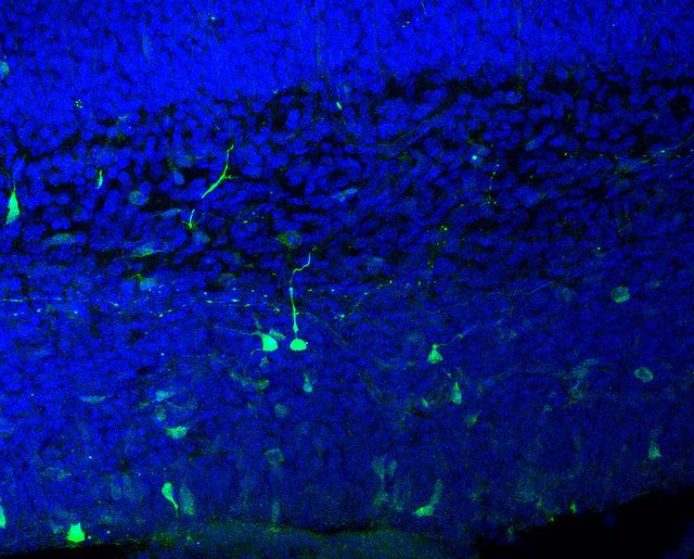
Led by senior author Dr. Jeff Lichtman, a professor of molecular and cellular biology at Harvard University, the inventors demonstrated their new automatic tape-collecting ultramicrotome (ATUM) on the brain of an adult mouse. To put it simply, what they did was cut the brain in slices, take a picture of each slice, and then put the images back together to form a 3D image on a computer, where it could be analyzed. This allowed them to create a detailed description of the brain.
In practical terms, the ATUM, which took two years to make, works like this: a diamond knife cuts sections from the brain at an ultrathin 29 nanometers. Immediately after they are cut, the sections are retrieved on a continuous conveyor belt. The pulling motion and adhesive tape of the conveyor belt force the captured sections to lie flat. Next, the tape is cut into strips and placed on silicon wafers that are photographed. Finally, the images are gathered together on a computer to form a completed picture of the brain. To make better sense of each image, the scientists used a special computer program to assign different colors as a way to piece apart each “object” (for example, neurons, glial cells, and blood vessel cells).
This video, courtesy of Cell and YouTube, explains this is in greater detail.
Even though this team of scientists is working with a minuscule area of the brain, one heart-stopping problem exists: a millimeter cubed, at nanoscale resolution, requires about two million gigabytes of computer data.
“Interestingly, the amount of data is close to what is currently transferred over the internet yearly," said Kasthuri. And, by comparison, the entire human genome is about three to five gigabytes. In short, the data storage demands for this research have never yet been encountered. However, just like genome sequencing, the researchers expect technology to continue to advance, thus ending these difficulties and lowering expenses over time.
The research was supported by, among others, the National Institutes of Health, DARPA (Defense Advanced Research Projects Agency), Intel, and Google, among others. This roster suggests the bandwidth of future practical applications for the technology is expected to be exceptional.
Source: Kasthuri N, Hayworth KJ, Berger DR, et al. Saturated Reconstruction of a Volume of Neocortex. Cell. 2015.
Published by Medicaldaily.com


