


In Living Color: GE 4D Heart Scans Are Like Nothing You've Ever Seen Before, May Help Doctors Save Lives
Earlier this year, GE Healthcare amazed us with the release of its high-resolution 3D CT scans. These realistic photographs allowed doctors to see inside the human body quite literally in living color. Now, GE’s latest body scanners instead use ultrasound technology to focus on capturing breathtaking shots of one particular muscle: the heart.
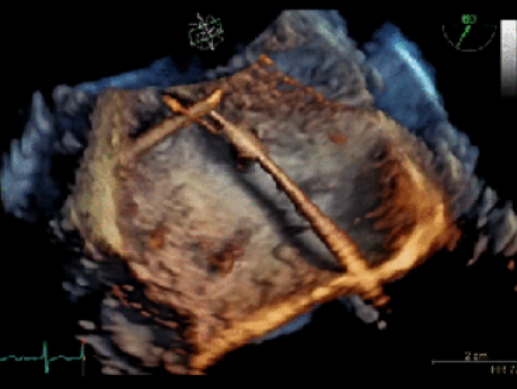
Ultrasound scanning, also known as sonography, uses high-frequency waves to create an image. They are used to help doctors to see what is occurring in a patient’s body in real time. It is a non-invasive test which allows doctors to both diagnose and treat patients.
The GE 4D scans work in a similar way but instead of using the traditional technology to create the realistic moving representations of the human heart, GE used a newly developed software known as cSound.
According to GE, cSound software is so powerful that it can compute a DVD’s worth of data in only a single second. This technology is able to not only process image pixels at an impressive rate, but also carefully choose only the best pixels to add to the final product. cSound software also has built-in color maps that color-code different tissues in the heart to make it even easier to view, Engadget reported.
“It’s like opening the chest and seeing the heart beating,” said Bijoy Khandheria, a cardiologist at Aurora St. Luke’s Medical Center, which is the first hospital to use the technology on patients, Time reported.
GE’s release of 4D images of the heart mark its attempt to tap into the U.S. heart health industry. According to the Centers for Disease Control and Prevention, about 5.1 million people in the U.S. have heart failure. Health services related to treating heart failure, including medications and missed days out of work, are estimated to cost the U.S. an estimated $32 billion each year. According to Engadget, GE believes this technology could help to shave some of the costs of heart-related diseases by eliminating the need for additional testing.
This would not be the first time that GE attempted to tap into a specific market with impressively realistic 4D images. In 2014, GE revealed its 4D ultrasound scans, which had four times the ultrasound pathway, 10 times the data transfer, and four times the processing power. The combination of these resulted in the most realistic images possible of life still inside the womb.
Correction: An original version of this story incorrectly described the GE heart scans as being the result of CT scanning technology. It has now been amended to read that they are obtained via ultrasound.


