


New Stem Cell Treatment May Be The Key To Reversing Age-Related Vision Loss

There could be a new treatment for vision loss on the horizon, thanks to embryonic stem cells and University of Montreal Professor Gilbert Bernier.
The researcher published findings Tuesday in the journal Development that detailed a new method of transplanting photoreceptors in the eyes. The transplantations offer a promising new treatment for a common form of vision loss called age-related macular degeneration (AMRD).
AMRD: How It Works
AMRD affects more than three million people in the U.S. every year and currently has no cure. It’s also the leading cause of vision loss among those 50 and older.
The common condition is caused by the degeneration of cells in the macula, a small but critical area of the retina at the back of the eye. The macula houses the retinal pigment ethelium (RPE), a tissue responsible for maintaining and repairing visual cells in the retina, and destroying the cells that are too worn out.
Cones, the cells responsible for color vision, are the photoreceptors affected by AMRD. We’re born with a fixed number of cones, and when they’re not maintained and repaired as much as they need to be, waste accumulates and forms deposits.
This causes gradual vision loss and is often first noticed as a blurred area near the center of vision. Over time, colors may not appear as brightly as they used to, and blank spots may pop up around a person’s central vision. Though ARMD does not lead to complete blindness, the vision loss can interfere with everyday activities and quality of life.
A New Approach
Bernier’s new treatment for ARMD has been a long time coming — he has been interested in the genes that code for the retina during embryonic production since completing his PhD in 1997. “I developed the idea that there was a natural molecule that must exist and be capable of forcing embryonic stem cells into becoming cones,” he said in a statement.
Bernier’s analytical approach led him to the idea of a little-known protein called COCO, described as a “recombinational” human molecule that’s expressed during the development of photoreceptors. Several years of research allowed him to isolate just how COCO’s molecular pathways are involved in photoreceptor development; he discovered that it systematically blocks all pathways that lead to the differentiation of other retinal cells. This discovery that allowed Bernier to begin producing photoreceptors.
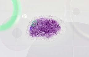
“Our method has the capacity to differentiate 80 percent of the stem cells into pure cones,” Dr. Bernier explained. “Within 45 days, the cones that we allowed to grow toward confluence spontaneously formed organized retinal tissue that was 150 microns thick. This has never been achieved before.”
To be sure the technique worked, Bernier experimented on mice, injecting clusters of the retinal cells into their eyes. The transplanted photoreceptors worked as they were supposed to, naturally migrating within the retina of their host.
“Cone transplant represents a therapeutic solution for retinal pathologies caused by the degeneration of photoreceptor cells,” Bernier said. “To date, it has been difficult to obtain great qualities of human cones.”
This stem cell driven treatment offers a solution to the problem, lending hope that treatments can be developed for currently incurable diseases like ARMD. Bernier acknowledges that the research has a long way to go before human clinical trials occur, but that in theory, the treatment will eventually be able to treat countless patients. Aside from the clinical application, the research could also aid scientists in creating models of human retinal degenerative diseases based directly on a patient’s own stem cell tissue.
Source: Zhou S, Flamier A, Abdouh M, Tetreault N, Barabino A, Wadwha S, Bernier G. Differentiation of human embryonic stem cells into cone photoreceptors through simultaneous inhibition of BMP, TGFB and Wnt signaling. Development. 2015.
Published by Medicaldaily.com


