


Personalized Brain Mapping: Diffusion Tensor Imaging Limits Tumor Surgery Risk
![See-Through Brain Allows Researchers To Study Brain Disease [VIDEO]](https://d.medicaldaily.com/en/full/226690/see-through-brain-allows-researchers-study-brain-disease-video.png?w=725&f=a17954c7d8c894111405f33074968c58)
Using a personalized brain mapping technique called diffusion tensor imaging (DTI), neurosurgeons can tailor brain surgeries so specifically that they can remove tumors without damaging parts of the brain responsible for key functions like speech, vision, and movement.
Researchers from the University of Pennsylvania's Perelman School of Medicine reviewed recent neurosurgery research on the personalized brain mapping method, and found that the visualization of white matter connections in the brain during brain tumor surgeries can significantly improve accuracy while limiting collateral damage.
"We can view the brain from the inside out now, with 3D images detailing connectivity within the brain, making a virtual intraoperative map," said senior author Dr. Steven Brem, co-director of the Penn Brain Tumor Center, in a news release.
He added that "we now have such detail about each individual's brain tumor -- combining diffusion tensor imaging and advanced imaging with the entire personalized diagnostics analysis available for all brain tumor patients," that each brain tumor surgery can be tailored to the unique neurophysiology of the patient.
The review, published in the journal Neurosurgical Focus, found that in a group of patients with high-grade brain tumors, the personalized brain mapping technique extended survival after surgery to about 21.2 months. Members of a control group where DTI was not used, on the other hand, survived to an average of 14 months after the surgery.
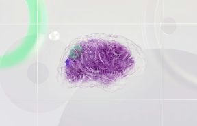
Diffusion tensor imaging is a type of magnetic resonance imaging (MRI) that tracks the movement and direction of water molecules along white matter tracts in the brain. The resulting 3D model of the white matter fibers is called a "diffusion tensor," and provides a color-coded map that literally displays how the brain's neurons are wired.
"The DTI images can be overlaid with structural and functional MRI images, providing a hybrid map showing topography layered with a road map," said Dr. Kalil Abdullah, lead author of the paper.
"This rendering gives us increased clarity to visualize important white matter tracts in the brain and adapt our surgical approaches to each person's case. Rather than focusing on solely taking the tumor out, we can avoid damage to healthy tissue and preserve important pathways responsible for speech, vision and motor function. "
The study authors write that diffusion tensor imaging's usefulness in personalized brain mapping for tumor surgeries makes it a key technique for the success of larger brain mapping projects, like the Human Connectome Project funded by the National Institutes of Health, and likely for the Obama administration's recently announced Brain Activity Map (BAM) project as well.
According to the release, the researchers are translating their findings to standard surgical practice. They hope to use personalized brain mapping with diffusion tensor imaging to further define how language deficits are defined by neural dysfunction before tumor surgery, and how they change or improve after treatment.
Published by Medicaldaily.com


