


Watch How A Virus Particle Enters A Cell In 3D
Sick of looking at chubby penguins, scraggly lemurs, and slow elephants? Tired of judgmental cats, useless fish, and parakeets that just sit there? You’re in luck.
This week, nature videography jumped up a notch with a new video from Princeton University showing a virus-like particle breaking into a cell. The project, led by postdoctoral researcher Kevin Welsher, aims to further the current understanding of biological interactions on the nanoscale. It also offers a glimpse into the life of a profoundly strange organism that, depending on the preferred definition, may not even be alive.
The unprecedented footage, which was shot in 3D, shows a nanoparticle (the dot) approach a cell (the green blob) and attempt to penetrate into the nucleus (the red blob inside the green blob). “Following the motion of the particle allowed us to trace very fine structures with a precision of about 10 nanometers, which typically is only available with an electron microscope,” Welsher explained.
In laymen’s terms, “very fine” would be an understatement. The camera is tracking a particle with a diameter of about 100 nanometers, or about 0.0001 millimeter. The cells, which belong to a class of skin cells called fibroblasts, are about 300 times larger. The entire scene takes place in the blink of an eye and could easily fit inside a human egg cell.
“What Kevin has done that is really different is that he can capture a three-dimensional view of a virus-sized particle attacking a living cell, whereas electron microscopy is in two-dimensions and on dead cells,” Haw Yang associate professor of chemistry and senior author of an accompanying study, said in a press release. “This gives us a completely new level of understanding.”
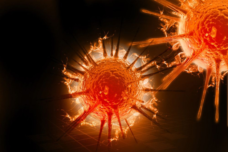
The particle is not really a virus particle, but a synthetic agent studded with protein segments derived the HIV-1 virus. Still, what we’re seeing is an incredibly intimate depiction of a day in the life of influenza, ebola, rabies, and other ills. These pathogens are all comprised of tiny, tiny particles called virions that penetrate the cell membrane and hijack the nucleus. By feeding host cells its own genetic material, the virus reduces the nuclei to tiny “virus factories” that spit out copies of virions until they literally burst.

Welsher’s footage was captured using two cameras — one for the particle itself, and another for the cells in the background. Besides helping virologists, the imaging technique may have tremendous benefits for drug development and delivery.
“We believe this will impact the study of how nanoparticles can deliver medicines to cells, potentially leading to some new lines of defense in antiviral therapies,” the researchers told reporters. “For basic research, there are a number of questions that can now be explored, such as how a cell surface receptor interacts with a viral particle or with a drug.”
Source: Welsher K, Yang H. Multi-resolution 3D visualization of the early stages of cellular uptake of peptide-coated nanoparticles. Nature Nanotechnology. 2014.
Published by Medicaldaily.com


