


It’s Congenital Viruses’ Effect On Neural Stem Cells That Causes Virus To Wreak Havoc On Fetal Brain Development
With the rise of Zika virus, there has been a surge of public interest in congenital, or mother-to-unborn child, transmission. Transmitting a disease in this way can cause abnormal developments in the fetus, most notably in the brain. Aside from Zika, viruses like HIV, rubella, measles, and cytomegalovirus (HCMV) are all capable of congenital transmission. Recently, a group of researchers developed their own model of infection to see exactly how viruses affect fetal brain development. They found that viruses can delay or even prevent the differentiation of stem cells into mature brain cells by activating a specific signaling pathway.
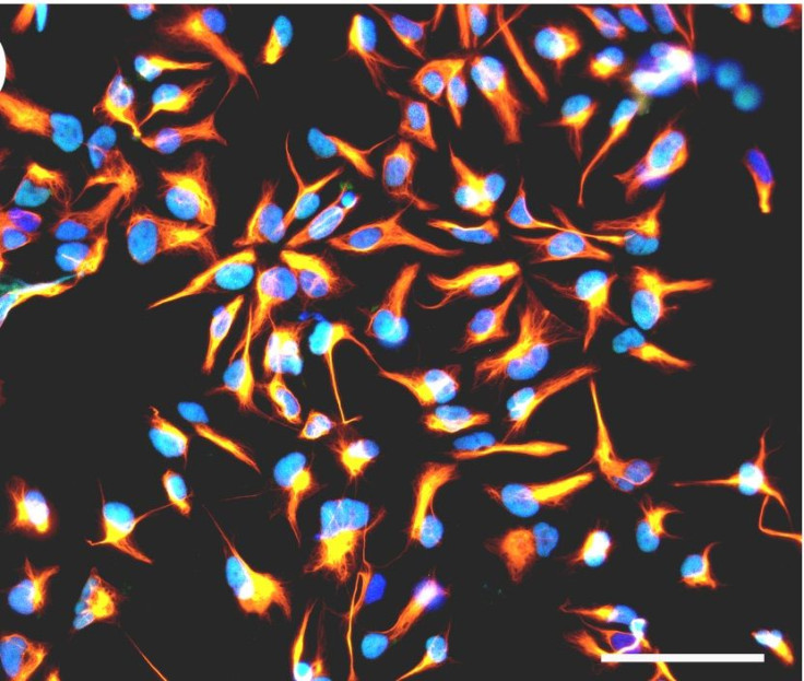
Stephane Chavanas, of the Universite de Toulouse in France, led a group of colleagues in developing an infection model based on human neural stem cells that normally produce neurons at a rapid rate. They observed that HCMV infection significantly reduced the number of neurons the stem cells could generate. They then dug further to uncover the possible mechanisms that allowed this to occur, and looked specifically at how the infection affected peroxisome proliferator-activated receptor gamma (PPARg), a protein critical to many cellular processes, including brain development.
Their study found that HCMV increased PPARg activity and overall levels in infected stem cells compared to uninfected cells. A known activator of PPARg, called 9-HODE, was more often present in infected cells as well. The infected cells also affected surrounding healthy cells — when liquid deposits from infected cells made contact with their healthier brethren, these too began to display elevated PPARg levels.
The scientists then worked to investigate the role PPARg played in stunting neuronal differentiation, and saw that healthy stem cells exposed to 9-HODE yielded lower rates of differentiation. They saw the same effects when activating PPARG in uninfected stem cells in the lab, too. “PPARg is sufficient to impair neuronal differentiation of human neuronal stem cells, even without infection,” the researchers wrote in the study.
Reversing the process, the researchers treated HCMV-infected stem cells with a drug that inhibits PPARg, and found that it restored the normal rate of neural cell production, backing up the link between PPARg and slower differentiation.
In the final part of the study, the team investigated PPARg expression in brain samples from real fetuses — 20 samples from aborted fetuses with congenital HCMV infection, and four samples from uninfected control fetuses. In the infected brain samples, they found PPARg was present in the central nucleus of cells in brain regions where healthy, active neuron production normally occurs. They observed no such thing in the healthy samples.
The researchers explained that human stem cells were an invaluable tool for modeling the correlates of HCMV infection. “This cell platform may probably be extended to other viral pathologies of the central nervous system,” they wrote, citing Zika as an example. They concluded that their findings “reveal a key role for PPARg in neurogenesis and in the pathophysiology of HCMV congenital infection, and… pave the way to the identification of PPARg gene targets in the infected brain.”
Source: Rolland M, Li X, Sellier Y, Martin H, Perez-Berezo T, Rauwel B, et al. PPARg Is Activated during Cogenital Cytomegalovirus Infection and Inhabits Neurogenesis from Human Neural Stem Cells. PLOS One. 2016.
Published by Medicaldaily.com


