


Tiny Lab-Grown 3D Brains Help Scientists Study How Zika Virus Damages Stem Cells In Brain

After months of research, the Centers for Disease Control and Prevention finally announced that there was enough evidence to conclude Zika virus does indeed cause microcephaly and other fetal brain defects. Now, scientists are delving into how exactly the virus causes these brain disorders — and they’re using tiny lab-grown brains to do so.
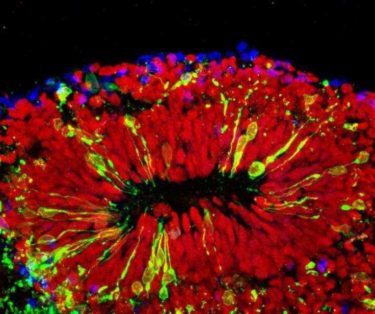
In a new study, researchers out of Johns Hopkins Medicine have found that Zika virus infects specialized stem cells in the brain’s outer layer, known as the cortex, contributing to damage linked to microcephaly and other fetal brain defects. Initially, the mini brains were created by a group of high school students who had developed 3D-printing technology during a summer research internship. Building tiny brains from this technology allowed the researchers to examine brain development and brain diseases on a detailed level.
“We have been working for three years to develop a better research model of brain development, and it’s fortunate we can now use this one to shed light on the major public health crisis posed by Zika infections,” said Hongjun Song, professor of neurology and neuroscience at the Johns Hopkins University School of Medicine’s Institute for Cell Engineering, in a press release. “This more realistic, 3D model confirms what we suspected based on what we saw in a two-dimensional cell culture: that Zika causes microcephaly — abnormally small brains and heads — mainly by attacking the neural progenitor cells that build the brain and turning them into virus factories.”
Mini brains are some of many tiny organs that have been grown in the lab to help researchers study the development of diseases on a level they could never do in patients. Recently, researchers used stem cell-grown mini brains to learn more about the mechanisms and development of autism spectrum disorder. Stem cells have also allowed us to grow mini retinas, kidneys, and other organs.
The latest study mainly focused on how Zika virus affected the brains of babies still in development, as scientists scramble to find ways to prevent or defeat the disease. On top of causing microcephaly — a severe brain abnormality in which babies are born missing part of their brain — Zika has been linked to Guillain-Barré Disease, which causes temporary paralysis, and various other disorders.
“One thing the mini brains allowed us to do was to model the effects of Zika virus exposure during different stages of pregnancy,” said Guo-li Ming, another researcher on the study, in the press release. “If infection occurred very early in development, the virus mostly infected the mini brains’ neural progenitor cells, and the effects were very severe. After a while, the mini brains would stop growing and disintegrate.” Even if the virus was introduced to the brain during the second trimester of the simulated pregnancy, it attacked the neural progenitor cells in addition to some other neurons. The researchers found that “growth was slower, and the cortex was thinner than in non-infected brains,” Ming noted.
In addition to studying disease development, the mini brains will allow the researchers to test out new therapies and drugs — the next steps in their research.
Source: Qian X, Nguyen H, Song M, Hadiono C, Ogden S, Hammack C. Brain-Region-Specific Organoids Using Mini-bioreactors for Modeling ZIKV Exposure. Cell. 2016.


