


3D Printing And Congenital Heart Disease: Heart Models Could Be Used To Plan Treatment
About 36,000 children born each year in the United States will come out the womb with a heart defect, known as congenital heart disease. Essentially, parts of these babies’ hearts haven’t developed properly — from the valves to nearby arteries or veins — causing symptoms like rapid breathing; a bluish tint in skin, lips, and fingernails; fatigue, and poor blood circulation. Treating these children can be expensive and emotionally tiring, as it involves highly specialized care. A new study, however, finds that 3D-printed hearts may eliminate the need for surgery, thus reducing costs.
“Children with congenital heart disease often need up to four open heart surgeries at different times of life,” biomedical engineer Dr. Peter Verschueren said in a press release. “The 3D-printed copy of the heart could reduce this to one or two, because doctors can choose and practice the best interventional approach and device beforehand. This will avoid children spending months in intensive care.”
3D printing has emerged as a useful tool in everything from aerospace to medical sciences. Just this year, scientists successfully created blood vessels using the sugar-based molecule agarose as a mold, and enclosed it in hydrogel, a gelatin-like substance. A team of researchers from the University of Louisville are also in the midst of developing a 3D-printed heart that may one day be transplanted — so far, they’ve only developed two-ventricle cylinders tiny enough for a mouse.
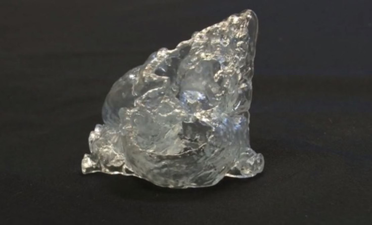
Verschueren’s 3D-printed hearts may not soon be transplanted, either, but they will give doctors a better idea of potential treatments. That’s because most current treatments are based on assessments from an image of the heart. By going 3D, the researchers will have a model of each patient’s unique, defected heart, which will even mimic beating. “3D models can be used to discuss the intervention with the medical team, patients, and, in the case of congenital heart defects, with parents,” said Helen O’Grady, another biomedical engineer, in the release. “It helps everyone affected to better understand what the procedure will involve.”
Most kids born with congenital heart defects go on to live relatively normal lives, albeit with some limitations, such as the ability to exercise. They’re also prone to heart failure and developmental delays in both motor function and learning. Some of these delays can be attributed to defects, however, other delays will probably come from time taken away from school to undergo surgery. It’s cases like these that make the 3D hearts useful, by allowing kids to live more normal lives.
Source: Verscheuren P, et al. At the European Association of Cardiovascular Imaging’s EuroEcho-Imaging 2014 annual meeting. 2014.
Published by Medicaldaily.com


