


AI Can Now Diagnose Tumor In Just 2 Minutes
The complex, difficult and time-consuming task of diagnosing brain tumors from tissue samples extracted during surgery stands to become a relic of the past with the advent of a new technology based on artificial intelligence (AI) that can do this job in about two minutes with an accuracy of 95 percent.
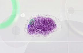
This powerful new AI-based tech optical imaging technique is called "stimulated Raman histology" (SRH), which works in conjunction with an artificially intelligent deep neural network. The system was developed by New York University neurosurgeons Dr. Daniel Orringer and Dr. Todd Hollon along with 35 colleagues as co-authors of the study.
SRH is a technology recently developed at the University of Michigan’s health system, which rapidly produces images of tumor tissue. It uses scattered laser light to illuminate features not normally visible to human eyes in standard imaging techniques. It works like this. During surgery, images acquired using SRH are evaluated by the AI algorithm. This super quick code needs less than 150 seconds to make its assessment compared to the 20 to 30 minutes required by human neuropathologists.
“This is the first prospective trial evaluating the use of artificial intelligence in the operating room,” Dr. Hollon said. “We have executed clinical translation of an AI-based workflow.”
In a published study explaining SRH, the authors “demonstrated how combining SRH with deep learning can be employed to rapidly predict intraoperative brain tumor diagnosis."
Even more useful is the AI is also capable of detecting features in the biopsies not visible to the human eye -- even a trained human eye such as those of a pathologist.
“As surgeons, we’re limited to acting on what we can see; this technology allows us to see what would otherwise be invisible, to improve speed and accuracy in the [operating room], and reduce the risk of misdiagnosis,” Dr. Orringer, senior author of the study, said. “With this imaging technology, cancer operations are safer and more effective than ever before.”
To develop the deep neural network needed by the AI, Dr. Hollon, the lead author, trained a convolutional neural network on 2.5 million images taken from 415 patients. The AI became so smart it could categorize tissues into any of 13 common forms of brain tumors. These include malignant glioma, metastatic tumors, diffuse astrocytoma, lymphoma and meningioma.
SRH was then tested in a clinical trial involving 278 brain tumor and epilepsy patients. The test was conducted at three different medical institutions to evaluate the efficacy of the system. Results showed the AI correctly identified the tumor 94.6 percent of the time. On the other hand, human neuropathologists were accurate 93.9 percent of the time.

Surprisingly, errors made by humans were different from those of the AI. The study said this discrepancy is actually good news because it suggests the nature of the AI’s mistakes can be accounted for and corrected in the future. The result is an even more accurate system.
“SRH will revolutionize the field of neuropathology by improving decision-making during surgery and providing expert-level assessment in the hospitals where trained neuropathologists are not available,” Matija Snuderl, co-author of the study and an associate professor at NYU Grossman School of Medicine, said.
Since many of the histologic features seen in brain tumors are also seen in other forms of cancer, SRH might eventually be used in other fields and surgeries. It might find use in dermatology, gynecology, breast surgery, and head and neck surgery, according to the study.
Published by Medicaldaily.com


