


AI vs. Humans: New Tech Spots Lung Cancer Better Than Doctors
Early detection of lung cancer is critical for both stopping the spread of tumors and improving patient outcomes, and a new study suggests artificial intelligence (AI) can do this job a lot more accurately than human doctors.
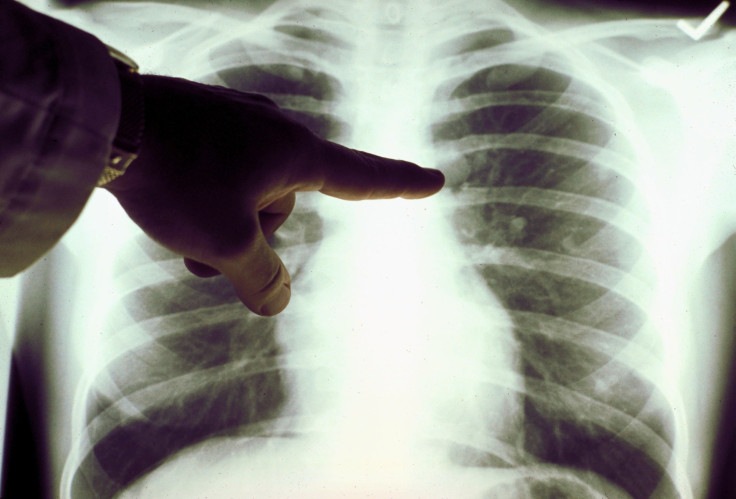
A recent study evaluating the use of a deep-learning algorithm to detect lung cancer accurately from computed tomography (CT) scans showed the AI can outperform human evaluation of CT scans.
Research also showed that low-dose CT (LDCT) reduces lung cancer deaths by up to 20 percent. There is a big drawback, however.
The high rate of false positives and false negatives resulting from doctors interpreting CT scans encumber the LDCT procedure. These errors delay the diagnosis of lung cancer until the disease has reached an advanced stage when it becomes too difficult to treat. New research stands to safeguard against these errors, however.
In the study, a group of scientists used AI techniques to detect lung tumors in LDCT scans.
Lung cancer causes some 160,000 deaths in the United States and is the leading cause of cancer-related death in the U.S. This means early detection is crucial for both stopping the spread of tumors and improving patient outcomes.
Researchers involved in the study resorted to a form of AI called deep learning to read 42,290 LDCT scans belonging to the Northwestern Medicine hospitals in Chicago. This deep-learning algorithm enables computers to learn by example.
The researchers were led by Daniel Tse from the Google Health Research group. Tse is also the corresponding author of the study, the findings of which were published in the journal, Nature Medicine.
Researchers trained the AI using a primary LDCT scan together with an earlier LDCT scan. Prior LDCT scans are useful because they can reveal an abnormal growth rate of lung nodules, thus indicating malignancy.
The study said the AI provided an "automated image evaluation system" that accurately predicted the malignancy of lung nodules without any human intervention. Researchers then compared the AI's evaluations with those of six board-certified U.S. radiologists with up to 20 years of clinical experience.
When prior LDCT scans were not available, the AI "model outperformed all six radiologists with absolute reductions of 11 percent in false positives and 5 percent in false negatives," said Tse. When previous imaging was available, the AI performed just as well as the radiologists.
"Radiologists generally examine hundreds of 2D images or 'slices' in a single CT scan, but this new machine learning system views the lungs in a huge, single 3D image," said study co-author Dr. Mozziyar Etemadi, a research assistant professor of anesthesiology at Northwestern University Feinberg School of Medicine in Chicago, in explaining why AI can outperform human evaluation.
He said AI working with 3D can be much more sensitive in its ability to detect early lung cancer than the human eye looking at 2D images. This capability can technically be regarded as “4D” because it not only looks at one CT scan but two (the current and prior scan) over time.
"Not only can we better diagnose someone with cancer, we can also say if someone doesn't have cancer, potentially saving them from an invasive, costly, and risky lung biopsy," said Dr. Etemadi.
The researchers caution that it’s first necessary to validate these results in larger cohorts.
Published by Medicaldaily.com


