Autism Could Be Detected Early In Infancy Using MRI Scans

Very little is known about the root causes of autism spectrum disorder (ASD), but it is known that some genetic factors may combine with environmental exposures to change the way the brain develops in young children. While there is no clinical treatment for the disorder, early intervention and education of children and parents can give affected children a greater chance of keeping up with other children in school and in social situations. To help this early diagnosis effort, researchers from the University of California, Davis MIND Institute have found that scans of the brains of infants may help to diagnose the condition earlier.
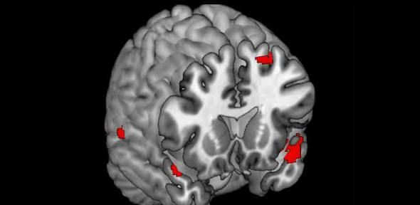
The study was originally designed to follow the brain growth trajectories of children and determine if there was a difference in brain growth of children later diagnosed with ASD. By backtracking the data to early time points, the researchers found that children who later develop ASD have excessive cerebrospinal fluid, the liquid that surrounds the brain and spinal cord, cushioning it and ferrying toxins away. The researchers also found that infants who later go on to be diagnosed with ASD have larger brains than other children, when they are infants. The test is performed by a safe and non-invasive MRI scan.
"This is the first report of an infant brain anomaly associated with autism that is detectable by using conventional structural MRI," said David Amaral, MIND Institute director of research and co-author of the study, in a press release. "This study raises the potential of developing a very early method of detecting autism spectrum disorder. Early detection is critical, because early intervention can decrease the cognitive and behavioral impairments associated with autism and may result in more positive long-term outcomes for the child," said Amaral.
The Study
The study was performed on 55 infants that were between the ages of six and 36 months. Of the 55 children, 33 had an older sibling already diagnosed with ASD and the remaining had no family history of ASD. The anomalous brain scans were seen at higher prevalence in children who had an older sibling diagnosed with ASD, which follows previous research showing that having an older sibling with ASD increases the risk of another child having it by 20 times. ASD is now diagnosed in one out of every 88 children in the U.S., according to the Centers for Disease Control and Prevention.
The MRI scans were performed three times for each child while they were asleep, so as to not use anesthesia or medications that may have affected the study results. At six months, the researchers assessed a multitude of behavioral traits in the children and gave parents questionnaires on their child's development. Of the high risk children, 45.5 percent were developing normally and 86 percent of the low risk babies were developing along normal benchmark paths.
The Results
At six to nine months of age, the researchers found that there was excessive cerebrospinal fluid in the extra-axial space above and surrounding the brain. The levels remained high through 18 to 24 months of age, and the amount of fluid in the space was proportional to the severity of ASD symptoms seen in children diagnosed. The fluid volume was 33 percent higher at 12 to 15 months, and 20 percent higher at 18 to 24 months for children eventually diagnosed with ASD compared to normal children.
The brain size of the infants eventually diagnosed with ASD was seen to be statistically larger than children who would not be diagnosed with the disease. Children who would go on to develop ASD symptoms had a seven-percent larger brain volume at 12 months, compared to children who develop normally. Both the extra cerebrospinal fluid and the larger brain size were detected far in advance of when behavioral tests would pick up signs of ASD. But the researchers do not know what is causing the increase in brain size and higher amount of cerebrospinal fluid.
"It is critical to understand how often this brain finding is present in children who do not develop autism, as well," said Sally Ozonoff, the vice chair for research and professor in UC Davis's Department of Psychiatry and Behavioral Sciences and co-author of the study.
What It Means
"For a biomarker to be useful in predicting autism outcomes, we want to be sure it does not produce an unacceptable level of false positives. If this finding of elevated extra-axial fluid is replicated in a larger sample of infants who develop autism, and it accurately distinguishes between infants who do not develop autism, it has the potential of becoming a noninvasive biomarker that would aid in early detection, and ultimately improve the long-term outcomes of these children through early intervention," said Mark Shen, UC Davis graduate student and the study's lead author.
Larger studies will be needed to determine if the small-scale data from 55 infants will be valid in scaled up population studies. Because of physical differences in brain neurons seen in people diagnosed with ASD, it is unlikely that the increased brain size and volume of fluid are causes of ASD, but rather may be a reaction to the development of a brain affected with ASD because of a physiological brain structure developmental difference compared to normally developing children's brains. If the results prove valid in larger studies, this could give doctors and parents an earlier biomarker that may indicate that the development of ASD is likely, helping to introduce interventional programs more quickly.
Source: Shen M, Nordahl C, Young G. Early brain enlargement and elevated extra-axial fluid in infants who develop autism spectrum disorder. Brain. 2013.
Published by Medicaldaily.com


