


Autism Speech Problems In Children Linked To Pleasure Disconnect In Brain
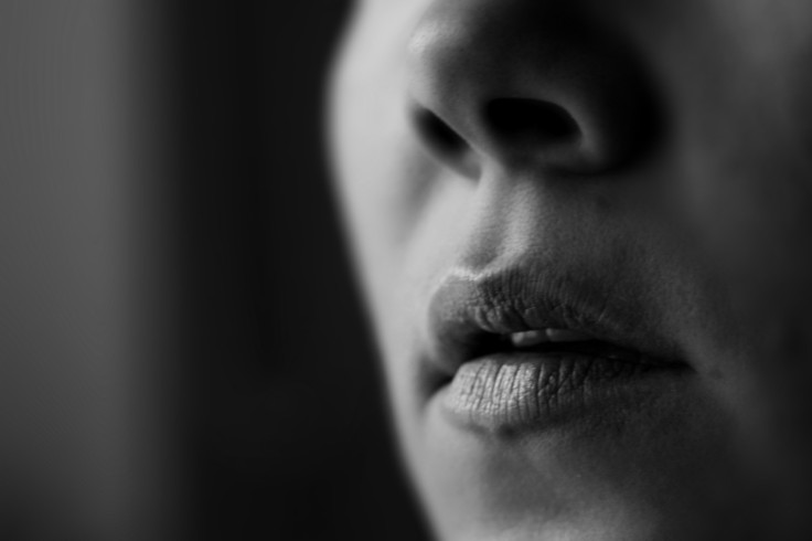
An essential quality of the human, speech serves to sooth and pleasure the child's brain, strengthening the bond between mother and child with release of oh-so-sweet oxytocin — a hormone closely linked to neurocognitive reward and general good vibe.
Within the brain of a child with autism spectrum disorder, however, atypical processing of the human voice may underpin deficits in ability to respond to such stimuli and the development of social behaviors crucial to full functioning. Alternatively, children and adults with the condition may experience a sensory deficit in which impaired processing of the acoustical features of sound precludes the brain's recognition of the human voice, leading to so-called autism speech problems.
Either way, researchers have begun to study the neural bases of voice processing with functional magnetic resonance imaging, looking for clues into the biology of autism. In adults with the condition, the brain fails to activate voice-selective regions of bilateral superior temporal cortex as compared to others, but not much is known about brain areas underlying voice processing and their links with systems distributed throughout the brain, those involving language, reward, and affective information processing.
Moreover, science has yet to investigate whether large-scale connectivity of the voice-selective temporal sulcus — associated with voice processing — is actually altered in the brains of people with autism, a glaring oversight given that connectivity problems elsewhere largely characterize the neurological basis of the condition.
In a small study published Monday in PNAS, researchers from Stanford University looked at those areas of the brain in 20 children with autism spectrum disorder and 19 others without the condition, who were of comparable age and intelligence. In the imaging, they found significant connectivity within a brain region called the basal ganglia in neurotypical children, as well as heightened connectivity among children with autism in the ventrolateral prefrontal cortex.
Other brain differences characterizing those with autism spectrum disorder was an absence of connectivity between the right hemisphere superior temporal cortex and several frontal, temporal, and parietal cortical regions, with connectivity limited to a fairly small region of superior temporal cortex. Neurotypical children also displayed greater connectivity in left hemisphere orbitofrontal cortex, bilateral ventromedial prefrontal cortex, and several subcortical brain structures.
"We demonstrate that childhood [Autism Spectrum Disorder] is associated with under-connectivity between voice-selective posterior temporal cortical regions and reward circuitry, providing important insights into the behavioral and clinical phenotype of abnormal speech and language processing observed in the disorder," the researchers wrote. "Critically, aberrant brain connectivity was associated with the severity of social communicative deficits in children with [the condition]. Our findings shed light on the neurobiological bases of one of the core deficits... by identifying key dysfunctional circuits associated with human voice processing."
Although small in scale, the study supports the social motivation theory of autism spectrum disorder, which posits that autism represents diminished social motivation in an individual, an unusual and profound lack of desire to seek acceptance and avoid rejection — the qualities typically voiced by one of the most social animals on the planet.
Source: Abramsa DA, Lyncha CJ, Chenga KM, et al. Underconnectivity Between Voice-Selective Cortexand Reward Circuitry In Children With Autism. PNAS. 2013.
Published by Medicaldaily.com


