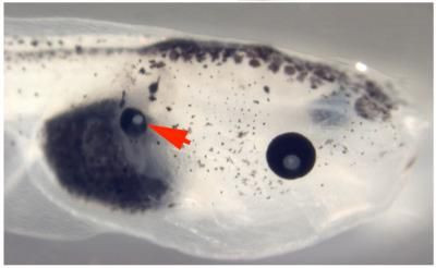
Bioelectric Change Prompts Tadpole Eye Growth in Back and Tail

Biologist at Tufts University were able to grow, for the first time ever, eyes outside of a tadpole’s head area and were able to control abnormal eyes.
Researchers achieved the feat by manipulating membrane voltage of cells in the tadpole's back and tail.
They changed the voltage gradient of cells to match that of normal eye cells, driving the cells located in the back and tail to develop into eyes.
"The hypothesis is that for every structure in the body there is a specific membrane voltage range that drives organogenesis," said lead author Vaibhav Pai, Ph.D.
"These were cells in regions that were never thought to be able to form eyes. This suggests that cells from anywhere in the body can be driven to form an eye."
Biology professor Michael Levin, Ph.D. of Tufts University said that in the field of biomedicine these findings break new ground because they identify an entirely new control mechanism that can be capitalized upon to induce the formation of complex organs for transplantation or regenerative medicine applications.
The researchers hope to control the incidence of abnormal eyes and repair such birth defects.
"These results reveal a new regulator of eye formation during development, and suggest novel approaches for the detection and repair of birth defects affecting the visual system," he said.
"Aside from the regenerative medicine applications of this new technique for eyes, this is a first step to cracking the bioelectric code."
Levin and his colleagues are pursuing further research to help them have a better understanding of how to control organ formation and treat visual birth defects.
Levin said that the findings will allow them to “have much better control of tissue and organ pattern formation in general. We are developing new applications of molecular bioelectricity in limb regeneration, brain repair, and synthetic biology."
Published by Medicaldaily.com


