Bone Marrow Cancer Spotted Anywhere In The Body With MRI Scans: Could New Technique Eliminate The Need For Painful Biopsies?

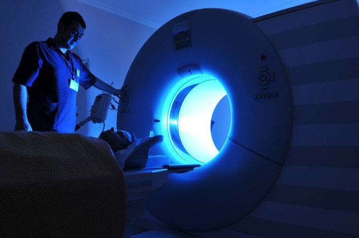
Patients with one of the most common types of blood cancer, myeloma, may soon get better, more efficient, and less painful treatment with a new magnetic resonance imaging (MRI) technique. Researchers who tested the technique say that one MRI scan was enough to pinpoint cancer in their patients’ bone marrow, anywhere in their body, without the need for biopsies or blood tests — the usual treatment.
Myeloma, also known as multiple myeloma, is a cancer that affects plasma cells, which are the white blood cells responsible for producing antibodies, according to the Mayo Clinic. As the cancer spreads, producing an abundance of abnormal plasma cells, the presence of abnormal antibodies in the blood also rises. This causes a range of health problems affecting everything from a person’s bones to their immune system to their red blood cell count.
Locating the cancer and seeing where it spreads can be a problem with current techniques, making treatment uncomfortable and sometimes painful for many. The new technique, known as a whole-body diffusion-weighted MRI scan, it can offer relief from repeated tests for an estimated 77,660 people. That’s because one scan could show where the cancer spread — if it spread — in each bone of the body, except for some parts of the skull. In 86 percent of cases, doctors were able to see whether patients were responding to treatment, while also seeing when they weren’t in 80 percent of cases.
“This is the first time we’ve been able to obtain information from all the bones in the entire body for myeloma in one scan without having to rely on individual bone X-rays,” said Nandita deSouza, professor of translational imaging at The Institute of Cancer Research in the UK, in a statement. “The results can be visualized immediately; we can look on the screen and see straight away where the cancer is and measure how severe it is.”
The Pain Of Bone Marrow Biospies
The MRI scan worked better than other methods for the 26 patients involved, the researchers said. Bone marrow biopsies, one of the stages of myeloma treatment, involve removing a bit of the soft tissue — the marrow — from inside the bone in order to determine whether the cancer has spread, or if a patient is responding to treatment. They can be uncomfortable, and painful, especially when anesthetic doesn’t work well.
Out of the patients who underwent an MRI scan, seven had undergone bone marrow biopsies just to learn that their samples couldn’t be used for analysis. Undergoing another biopsy could have further traumatized them, and there was no guarantee that findings would have been different. Other forms of treatment like X-rays and blood tests are time consuming, and their results are limited.
The MRI helped doctors get around all of these challenges while also helping them assess whether or not treatment was working. “Myeloma can affect bones anywhere in the body, which is why this study is so important,” said Dr. Faith Davies, a member of the Myeloma Targeted Treatment Team at the Institute of Cancer Research, in the statement. “We’ve shown that whole body MRI scans can accurately monitor how myeloma patients are responding to treatment, allowing doctors to make more informed decisions. With this new scan, if a treatment isn’t working, the patient can be moved onto new therapies that might be more effective much more quickly.”
Though the MRI technique is very promising, the researchers note that a much larger group of patients still need to be tested in order to prove that it works. “This research demonstrates how an advanced imaging technique could provide a whole-skeleton ‘snapshot’ to track the response of tumors in individual bones,” said Julia Frater, Cancer Research UK’s senior cancer information nurse, in the statement. “Finding kinder ways to monitor how patients respond to treatment is really important, particularly in the case of myeloma where taking bone marrow samples can be painful.”
Published by Medicaldaily.com


