


Breakthrough in IVF Could Double the Rate of Successful Births
In vitro fertilization (IVF) success rates may go up as a result of a new technique that will help determine which embryos are most viable for live birth, according to new research.
Scientists at the CARE Fertility Group in Nottingham England developed the technique in which time-lapse photographs are taken during the first five days of the IVF embryo's life. During this period, the embryo first appears as the blastocyst — a ball usually comprised of about 70-100 cells. Scientists discovered that if blastocyst took longer than about six hours to form, the embryo was likely to be carrying an abnormal number of chromosomes, also known as aneuploidy. The occurrence of aneuploidy has been shown to fail at initiating pregnancy, according to The Independent.
The average success rate for IVF is 30 to 35 percent for U.S. women under age 35, 25 percent for women aged 35 to 37, 15 to 20 percent for women aged 38 to 40, and 6 to 10 percent for women over 40 years old, according to the American Pregnancy Association.
"I believe it is the most exciting breakthrough we've had in probably 30 years," Professor Simon Fishel, managing director of CARE, said to The Independent. "We hope to see a paradigm shift in terms of IVF. It's a game changer for everybody to have such an uplift in birthrate. This is the beginning of something revelatory."
Although it is a preliminary study, the analysis examined 88 IVF embryos of 69 couples and found that the time-lapse technique could have improved the success rate of live births in this group from 39 percent to 61 percent. Scientists expect success rates as high as 78 percent once the technique is refined and applied to the wider population of infertile couples.
"Our work has shown that we can easily classify embryos into low or high risk of being chromosomally abnormal. This is important because in itself this is the largest single cause of IVF failure and miscarriage," Fishel said. "The beauty of this technology is that the information is provided by a non-invasive process. So far we have seen a 56 percent uplift compared to conventional technology, giving our patients the equivalent to a 78 percent live-birth rate."
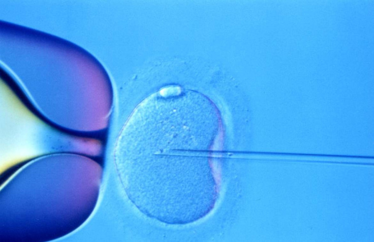
Normally, embryos undergo a biopsy in which samples are taken from the growing embryo manually, but with the time-lapse technique, they aren't touched. Instead, a camera takes pictures every 10 minutes without interfering with the embryo's development, The Independent reported.
"With time-lapse we have the ability to view more than 5,000 images over the same time period to observe and measure more closely each stage of division and growth," Alison Campbell, embryology director at CARE, said. "As a result of continuous monitoring we have demonstrated that delays at defined points indicate abnormal development."
Researchers, however, emphasize that more research must be done to compare the new techniques to existing ones.
"Unfortunately the study does not compare this exciting new approach with standard practice in embryology in which embryologists already look for the best embryos to place in the womb," Sue Avery, director of Birmingham Women's Fertility Clinic, said. "Until the new technique is compared to current practice we cannot know whether different embryos are being chosen."
Published by Medicaldaily.com


