


Catching Feelings: Traffic In Amygdala Neural Circuit Determines If We Process Emotions As Good Or Bad
The brain, our most complex organ, is an ever-active communication hub where cells continuously speak with other cells. Naturally, connections between neurons in the brain are key to the ability to process our many thoughts and emotions. In fact, scientists believe the origin of many if not all brain disorders, including depression, addiction, and post-traumatic stress disorder, may be a misrouting of information.
A new study explores the way two separate populations of collaborating cells within the amygdala, the emotion zone of our brain, contribute to our ability to assign feeling to particular events.
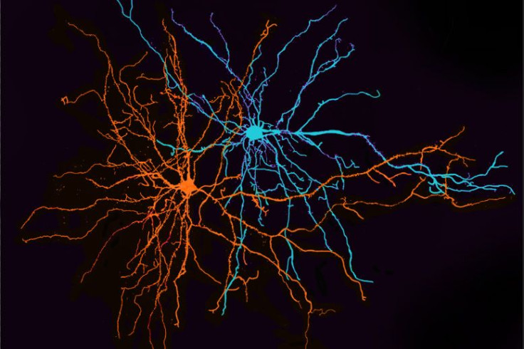
Though they intermingle, two groups of neurons in the amygdala will form parallel lanes to carry information about an experience to other cells; as the researchers explain, this traffic is what decides whether our emotional response to an experience is positive or negative. Led by Dr. Kay Tye, of MIT's Picower Institute for Learning and Memory, the team believes the reason these different neuron populations reside together within the amygdala is to allow for quick interactions among cells reacting to different inputs. Such speed is necessary, they hypothesize, when coordinating a response to urgent situations.
The current study is based on previous research performed by Tye's lab, where she and her colleagues first identified the two populations, or groups of functionally similar neurons in the amygdala. At that time, they learned how these separate groups of cells helped process both positive and negative emotions. One of these populations relays information to the nucleus accumbens, which plays a role in learning to seek rewarding experiences, while the other sends input to the centromedial amygdala, which scientists believe may turn our attention to important stimuli and influence how we react to situations.
Intrigued by this discovery, the researchers wanted to know more. They asked themselves, what exactly do these neurons do when an animal reacts to a frightening stimulus — or, for that matter, a pleasurable one? And so they designed the current experiment.
The Experiment
They began by tagging, in three groups of mice, each population of neurons. This is necessary because, unlike some areas of the brain, no topographical features separate the populations or limit how they move so they are intermingled and otherwise indistinguishable. Using a light-sensitive protein as a tag, the researchers labeled the cells communicating with the nucleus accumbens, the cells projecting to the centromedial amygdala, and a third group of cells connecting to the ventral hippocampus, which is involved in anxiety. Next, the researchers trained the mice to discriminate between two different Pavlovian sounds, one linked to sweet reward (sugar water) and the other to bitterness (quinine). Finally, they recorded electrical activity from each group of neurons as the mice encountered the two stimuli.
Analyzing the collected data, the scientists compared the brain’s anatomy (which neurons are connected) and its physiology (how neurons respond to input) and discovered a surprise.
The neurons within each labeled subpopulation did not all react the same way to each stimulus. Some responded to one stimulus, some to the other, and some responded to both. Some neurons became excited and others inhibited.
Despite these many differences, the researchers discovered overall patterns of behavior for each population. The neurons projecting to the ventral hippocampus appeared balanced, responding equally to both positive and negative cues. Among neurons projecting to the nucleus accumbens, the reward stimulus excited most, while bitterness drew no reaction. Finally, the opposite happened among neurons connecting to the central amygdala — the bitter cue excited most of these neurons, which remained unresponsive to the sweet reward.
These results suggest our brains process emotions at the level of small populations of cells or possibly even individual neurons. As Tye and her colleagues discovered, we have only scratched the surface and do not yet fully appreciate the complexities of our brains.
Source: Beyeler A, Namburi P, Glober GF, et al. Divergent Routing of Positive and Negative Information from the Amygdala during Memory Retrieval. Neuron. 2016.
Published by Medicaldaily.com


