


Glaucoma Breakthrough 2016: Early Detection and Experimental Drug Could Help With Vision Loss, Researchers Say
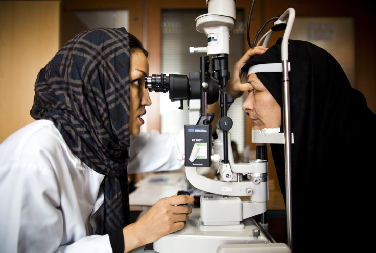
Researchers for the first time identified distinct characteristics of different types of glaucoma using optical coherence tomography (OCT) angiography by observing the disease in its earlier stages. The findings published Friday in the journal IOVS could help doctors diagnose the illness earlier, which could help slow down the loss of vision associated with glaucoma.
The disease, which is characterized by progressive loss of vision, occurs when the optic nerve connecting the eye to the brain is damaged due to pressure. OCT angiography, an advanced imaging system, can capture the movement of red blood cells in the blood vessels without having to use dye injections as is the case with traditional angiography.
Researchers from the Mount Sinai Hospital in New York and Rutgers New Jersey Medical School found that patients with glaucoma have poor blood flow compared with people who don’t have glaucoma. They observed 92 people between April and August 2015. All the patients were over 50 years of age and belonged to one of the three groups: primary open-angle or high-pressure glaucoma, normal-tension or low-pressure glaucoma, and no glaucoma.
On observing the patients’ blood vessels using OCT angiography, the researchers found that patients with glaucoma had different defects in the blood vessels depending on the type of glaucoma they had.
“This is the first time we have been able to identify certain characteristic patterns of blood flow that correspond to different types of glaucoma, which may allow us to identify certain forms of glaucoma in their early stages,” lead investigator Richard Rosen said in a statement. “The findings could lead to new therapeutic strategies to avoid progressive damage in glaucoma patients, and provide a new metric for monitoring early damage that eventually leads to vision loss.”
Researchers developed a new software that can analyze the images obtained using the OCT angiography. They found that in patients with high-pressure glaucoma, the pattern of blood flow loss was similar to optic nerve fiber loss. In patients with normal-tension glaucoma, the blood flow pattern tended to be more diffuse. The blood flow pattern in patients with normal-tension glaucoma was not very different from the one seen in open-angle glaucoma patients.
“Normal-tension glaucoma is often picked up late because it’s not recognized if patients don’t have high pressure, causing them to lose nerve fiber and blood supply before diagnosis,” said James C. Tsai, president of Mount Sinai’s New York Eye and Ear Infirmary. “This study points to vascular factors contributing to normal-tension glaucoma and we can use this to help identify ways to detect the disease earlier, and possibly prevent vision loss.”
Meanwhile, an experimental drug, developed by researchers led by Jeffrey Goldberg from the Byers Eye Institute at Stanford University, when implanted in the eye could stimulate activity and growth of the optic nerve.
BrightFocus Foundation, a nonprofit organization that funds glaucoma research, said Friday the NT-501 encapsulated cell therapy, or NT-501 ECT, could play a new role in fighting glaucoma. The drug is a capsule filled with genetically modified human cells. These cells secrete ciliary neurotrophic factor, which can stimulate nerve growth and activity. The Phase-2 clinical trial, which was launched recently, will observe subjects for two years looking for any change in their vision.
The drug targets the retinal ganglion cells in the eyes that carry light signals to the brain. Researchers believe that the drug might put a stop to damage to the retinal ganglion cells or improve existing vision.
“It will be an enormous step forward if either of these can be demonstrated,” Goldberg said in a statement. “We have no approved treatments that address the degeneration of the RGCs or their axons, so this is a huge unmet need.”
Published by Medicaldaily.com


