


How Do You Know If You Have Cancer? Fluorescent Protein May Illuminate Spread Of Cancer Cells
In an effort to better root out any remaining cancer cells, scientists from the University of Arkansas for Medical Sciences have developed a fluorescent protein tracking system that may potentially allow oncologists to stop cancer’s spread in real-time.
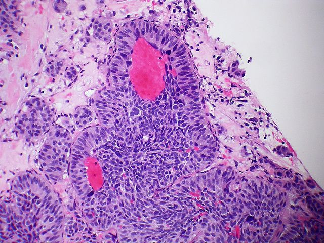
The new study, published in the journal Chemistry & Biology, adds to a growing body of evidence that suggests metastasis, the process of cancer cell growth, can be halted if scientists have a way to know where it’s happening. When cancer turns fatal, it is often the result of unchecked growth — to the extent that doctors can no longer operate on the patient without damaging surrounding structures.
In fact, metastasis is so threatening that it leads to up to 90 percent of all cancer deaths. That is why early treatment is ideal, as the cancer hasn’t yet had a chance to invade other tissues and replicate. But despite the known danger of allowing cancer to branch out, unrestricted, the advances that have been made still remain mostly experimental. Few resources exist that allow clinicians and oncologists to monitor cancer’s progress with confidence and stop it in time.
Senior author of the study Dr. Ekaterina Galanzha explains that the new technology monitors the real-time behavior of circulating cancer cells sent from a primary tumor. But more than that, it “allows for the labeling of just one circulating pathological cell among billions of other normal blood cells by ultrafast changing color of photosensitive proteins inside the cell in response to laser light,” she said in a statement.
The illumination works via a photoswitchable fluorescent protein, meaning the researchers have the ability to label individual cells using a particular color light. One wavelength turns the cells green, and another red. To label the cells, they use a thin violet light. Once all the desired cells are tracked and labeled, the team can reproduce them on a computer monitor to count them.
“The approach may give oncologists knowledge on how to intervene and stop circulating cancer cell dissemination that might prevent the development of metastasis,” Galanzha said.
The technique isn’t limited to cancer. Given the success of tracking specific fluorescent proteins, researchers should have the ability to trace any disease or bacterial infection that may course through people’s bodies — including “immune function, bacteremia, sepsis, and clotting,” the team wrote in their report. “In the future, this knowledge could help in developing advanced diagnostic techniques and individualized therapy.”
Source: Nedosekin D, Verkhusha V, Melerzanov A, Zharov V, Galanzha E. In Vivo Photoswitchable Flow Cytometry for Direct Tracking of Single Circulating Tumor Cells. Chemistry & Biology. 2014.
Published by Medicaldaily.com


