


New Way Found to Image Brain Tumors for Better Survival
Patients who undergo neurosurgery after being diagnosed with low-grade glioma, a type of brain cancer, are still at risk of developing a more malignant state of cancer and their odds of survival has been unknown until now.
Researchers from the University of California, San Francisco (UCSF) have developed methods to reveal a molecular marker in tissue samples from brain tumors that has been linked to better survival odds, providing doctors with a better way in monitoring their patients brain activity after surgery.
“If a tumor transforms to a higher grade, then it is important to use more aggressive treatments,” said Sarah Nelson, PhD, the Margaret Hart Surbeck Distinguished Professor in Advanced Imaging at UCSF and a professor in the Department of Radiology and Biomedical Imaging.
The authors explained that this new technique might help doctors better gauge cancer recurrence, make follow-up treatment decisions and assess how a patient responds to recommended treatments.
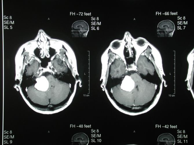
The researchers used MRI methods to obtain data from image-guided tissue samples from more than 50 patients with glioma that demonstrated the presence of a chemical called 2-HG, linked to mutations in a gene known as IDH1, the authors wrote.
But further research must be done, explained the authors, as the techniques must be refined to allow standard hospital MRI imaging to show 2-HG.
"Developing methods to obtain images in a clinical setting is an engineering challenge now," said Nelson
Published by Medicaldaily.com


