


The Picture Perfect Future of Medical Imaging
X-rays and low quality images are a thing of the past. In the world of medicine, new imaging technology provides dynamic pictures that are revolutionizing how doctors treat patients and diseases.
Medical imaging goes well beyond high-definition into the world of 3D. X-rays or even those ultrasound pictures of an infant are low-detail and in this day and age, medical imaging is letting doctors not just take photos but create 3D models of a patient or a disease. In just a few short years, these advances have shaped how surgery and disease treatment are performed.
The study outlining of these amazing advancements of medical imaging was led by Charl Botha, PhD, from the Delft University of Technology in the Netherlands. Not only has medical imagery evolved so rapidly in the last few years, the future is looking even brighter with even more advances in medical imaging yet to come.
Some familiar advances in medical imaging include magnetic resonance imaging (MRI) and computed tomography (CT) scanners. Magnetic resonance imaging (MRI) made looking at X-rays like watching a flickering black-and-white television thanks to their ability to produce high contrast images.
CT scans and MRI's have allowed for 3D models of a patient and new CT scanners can take enough image data in a second to create a 3D video of a beating heart, according to researchers. Surgery and cancer treatments also rely heavily on CT scans and MRI's, note researchers.
While clarity and the ability to distinguish between tissues and tumors, the images don't exactly look like how a human does in real life. That's about to change though as new imaging advances which focuses on the diffusion of water through the body and tissue and better lighting techniques create photo-realistic images of what's inside a human.
New imaging techniques involving water diffusion helps doctors examine otherwise difficult to image parts of the bodies such as muscle fibers. This technique can help doctors better understand the nervous systems and the ways our body moves.
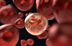
Other imaging advances have occurred on the molecular level, allowing researchers to gain a glimpse into the origins of a disease or a tumor.
These advances have helped doctors detect diseases much earlier than before and that will save countless lives in the future. While these advances are impressive, medical imaging has advanced even further when it comes to detailing the surface of objects.
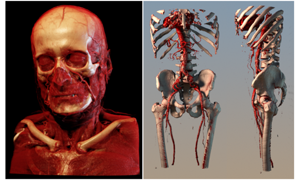
In the field of medical data topologically, images can depict realistic images of organs and let doctors understand the shape an organ, greatly aiding surgical procedures. Above all, these imaging advances have let doctors use lighting techniques that provide photo-realism. Tissue is a vibrant red and bones actually look like bones.
In the future, researchers hope imaging techniques can be developed to show the progress of disease as shown in multiple individuals or how it differs between different groups. Other imagery advances may be able to help create potential images of surgical outcomes.
Medical imaging is changing how doctors see patients but how doctors actually view the images have also changed. Tablet devices, such as an iPad, are providing portable methods that can improve a doctor's productivity. With photo-realistic images, molecular image and other developments, the future is in focus thanks to advances in medical imaging.


