


Conjoined Twins Separated: Doctors Disjoin Major Organs In 26-Hour Operation To Separate Twin Girls
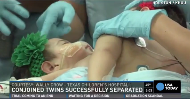
Doctors at Texas Children’s Hospital had their work cut out for them during a 26-hour operation to separate conjoined twins Knatalye Hope and Adeline Faith Mata. After spending their first 10 months of life conjoined at the chest and abdomen, Knatalye and Adeline underwent the grueling operation in which surgeons disjoined their chest wall, lungs, diaphragm, liver, intestines, colon, pelvis, and the lining of the heart, also known as the pericardial sac.
"This surgery was not without its challenges with the girls sharing several organ systems," Dr. Darrell Cass, pediatric surgeon and co-director of Texas Children's Fetal Center, told KHOU. "Our team has been preparing for this surgery for months, and we've done everything from working with our radiology experts to build a 3-D model of their organs, to conducting simulations of the actual separation surgery."
According to the American Pediatric Surgical Association, around 250 successful separations in which one or both twins survived over the long term have been recorded to date. The majority of these successful separations occurred within the past two decades thanks to medical innovations, including radiology imaging technology, surgical techniques, and anesthesia.
To separate Knatalye and Adeline, surgeons had to call in help from specialists in pediatric surgery, plastic surgery, cardiovascular surgery, urology, liver transplant surgery, orthopedic surgery, and pediatric gynecology.
Published by Medicaldaily.com


