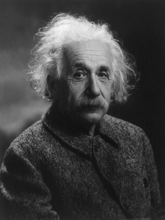
Rare Brain Photographs Show Einstein's Brain Was Unique

Einstein's extraordinary cognitive abilities came from certain structures in his brain that are different from those found in brains of other people, says a new study.
Dr. Thomas Harvey had conducted the autopsy on Albert Einstein at Princeton Hospital on April 18, 1955. He first cut the brain in 240 blocks and later sliced the blocks in 2,000 sections to study them under a microscope. Over time, he distributed these microscopic slides to many research facilities around the world.
Recently, The National Museum of Health and Medicine Chicago had launched an app that made it possible for the general public to see parts of Einstein's brain.
For the study, researchers analyzed 14 recently discovered photographs of Einstein's brain and compared them to 85 brains that were earlier studied. The photographs analyzed in the present study are held by the National Museum of Health and Medicine.
"Although the overall size and asymmetrical shape of Einstein's brain were normal, the prefrontal, somatosensory, primary motor, parietal, temporal and occipital cortices were extraordinary. These may have provided the neurological underpinnings for some of his visuospatial and mathematical abilities, for instance," Dean Falk, evolutionary anthropologist, Florida State University and lead author of the study.
Einstein's ability to play the violin would have come from unusual feature in the right somatosensory cortex that is associated with sensory information coming from the body. In Einstein's brain, the area that corresponds to left hand is expanded, Nature reported.
The study is published in the journal Brain.


