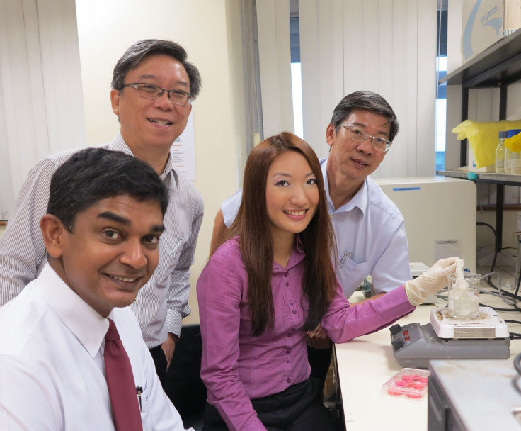
Scientists Engineer 3-D Tumor Model Using Silk For Cancer Drug Testing

As part of their research program on osteosarcoma, scientists at National University of Singapore (NUS) have engineered a highly realistic 3-D tumor in an effort to replicate the body’s internal conditions and allow for more precise measurements of drug activity. The key to their success? Silk.
The NUS team, comprised of researchers from the Department of Bioengineering as well as the Department of Orthopaedic Surgery, has been working on the tumor microenvironment for the better part of a decade. On Thursday, it was announced that their first 3-D reconstruction of a cancerous tumor had been successfully engineered in a pressurized bioreactor. The process requires cells to be implanted – or “seeded” – into artificial structures called scaffolds that promote cell growth by emulating biological conditions. The NUS team’s reconstruction of the osteosarcoma tumor relied on scaffolds made from silk – a fiber that appears ideal for both cell attachment and growth.
Until now, treatment activity within cancerous tumors has been notoriously hard to map, as current in-vitro testing cannot fully predict treatment efficiency within actual conditions. The reason is that laboratory testing largely uses 2-D cell culture systems that lack a number of structural features particular to the real, 3-D environment within the body. On the other hand, in-vivo methods – where research is focused on actual tumors in actual patients rather than artificial models – are unfeasible for comprehensive, large-scale biological experimentation.
The new, realistic 3-D model – the first ever created using silk scaffoldings – could represent the first step in bridging this troublesome gap. The team found that results obtained from their 3-D tumor, when compared to those of 2-D laboratory methods, were much closer approximations of results obtained from actual in-vivo samples. In addition, chemotherapeutic testing indicated that the tumor construct responded to doses within those measured in lab mice – a discovery that may reduce the need for live animal testing.
Associate professor Saminathan Suresh Nathan from the Department of Orthopaedic Surgery at the NUS Yong Loo Lin School of Medicine noted that the new method will eventually extend beyond osteosarcoma research, and that the discovery will have significant bearing on a variety of other treatment programs. Future developments will focus on implementing other aspects of the real-life microenvironment, such as oxygen levels, and eventually establishing a reliable, comprehensive platform for anticancer drug testing.
Published by Medicaldaily.com


