Learn resistance training basics with focus on proper form, compound lifts, and beginner weights to build strength safely through steady, progressive overload for lasting fitness results.
Discover how everyday housework boosts fitness. Learn how vacuuming, mopping, and gardening act as effective workouts that help you achieve daily exercise goals naturally.
Discover how small coffee and espresso tweaks can create heart healthy coffee habits and sleep friendly routines while keeping your daily caffeine enjoyment balanced and satisfying.
Discover how social media effects contribute to rising teen anxiety, supported by mental health studies, coping strategies, and parental tips for protecting adolescent well-being online.
Explore how the health effects of loneliness and social isolation are becoming a major public health risk, impacting physical and mental well-being across all ages globally.
Explore how wearable health technology in smartwatches tracks vital signs, spots early warning patterns, and supports smarter, more proactive health decisions in everyday life.
Explore how CRISPR gene editing is transforming genetic disease treatment, uncovering breakthroughs, safety advances, and the future possibilities of curing inherited disorders through DNA repair.
Artificial skin technology and electronic skin research enable prosthetics, robotics, and burn grafts to sense touch, temperature, and textures like real skin.
Discover how smart thermostats and health are connected by optimizing bedroom temperature to improve sleep quality, support easier breathing, and create a more comfortable, restorative sleep environment.
Boost fat loss and endurance with HIIT exercises using metabolic conditioning and the Tabata protocol, delivering fast, time-efficient workouts that maximize calorie burn and performance.
It’s reported that up to 70% of the population have sensitive skin. More alarming, allergies and skin sensitivities are known to affect millions of patients taking prescriptions every year.
Tech News: NIH Considers New Photon CT Scanner With Maximum Image Quality, Minimum Radiation Exposure
By
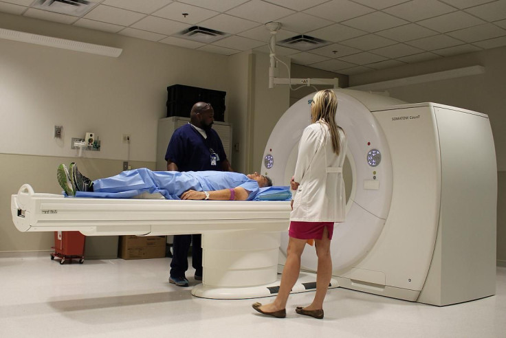
The NIH's new photon-counting detector CT scanner is expected to surpass the conventional machine by offering an enhanced look inside the body.Courtesy of the National Institutes of Health
Though it looks much the same on the outside, a new computerized tomography (CT) scanner is a whole new story when it comes to the inside — the inside of a patient, that is. A photon-counting detector CT scanner is being given a pilot run at the National Institutes of Health, where researchers investigate its use in a medical setting. The prototype technology from Siemens Healthcare is expected to surpass conventional CT scanning by providing an enhanced, more detailed look inside the body.
Best of all, the technology delivers only a minimum amount of radiation to patients lying in the scanner bed.
Why is the NIH looking into this new machine? The advanced technology increases both the resolution and contrasts available for analysis, so it may be instrumental in improving diagnosis, the scientists say in a statement to the press. Among its benefits:
Doctors should be able to see selected areas of the body in greater detail and with anatomic precision.
By using new contrast agents, different materials in the body will be displayed in different colors for faster diagnosis and precision.
The technology can identify and characterize tumors, plaques, or vessels that are smaller than half a millimeter.
The new CT scanner also can identify soft tissues such as proteins, tendons, or collagen, which are difficult, if not impossible, to differentiate with the current equipment.
The image below shows you the color and resolution captured by the scanner.
Scanned ImageCourtesy of the NIH
Algorithms and Protocols
In the study and treatment of disease, surgery is often viewed as the last option. CT scanning is one way that doctors can examine the body’s internal features without resorting to a patient undergoing the knife. The scan works by taking X-ray images from different angles and then using an algorithm and computer processing to combine the series of images and create cross-sectional pictures, or slices, of the bones, blood vessels, and soft tissues inside your body. These scanned images can be used to diagnose injuries as well as disease and frequently doctors use them to plan surgeries or other medical treatments.
The NIH Clinical Center, which sees thousands of patients every year, is one of three sites in the world to use this technology. More than 45 volunteers enrolled in a research study of the equipment. Over the next five years, Dr. David Bluemke and his colleagues in the department of radiology and imaging sciences will develop algorithms and scan protocols in the hopes of improving diagnosis, screening, and treatment planning for cancer and cardiovascular disease.