


A Transparent Fish Shows How Meningitis Moves Through The Body And Attacks The Brain
In a new study released by Duke University Medical Center, researchers created a video showing how Cryptococcal meningitis takes over the brain. Using a transparent fish, the resulting images look like something straight out of a sci-fi movie teaser, but they will give scientists further insight into how meningitis works in the body. Published in mBio, their findings bring them one step closer to helping the 600,000 people worldwide who die of this disease.
According to the study, Cryptococcus is a form of fungi we breathe in almost every day. For most of us with healthy immune systems, the fungi does little harm. However, those who suffer from conditions that compromise the immune system, like HIV, are at greater risk of the infection taking hold and becoming deadly.
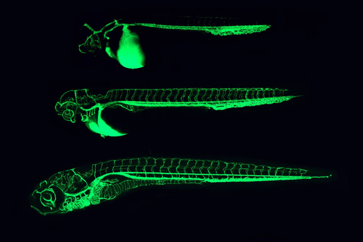
In order to develop effective medications, the researchers knew it was key to understand how the infection moves from the lungs into the bloodstream, and eventually into the brain. Microscopic zebrafish larvae seemed like a good specimen to experiment on; having clear bodies, researchers were able to inject the organism with the fungi and see how it moves.
“What’s impressive is that, unlike in a mouse or a rabbit, you can actually see the organism producing disease in the live animal,” said Dr. John R. Perfect, chief of the division of infectious diseases at Duke University School of Medicine, in a recent press release. “Day-by-day, it’s growing and moving throughout the body. You can’t see this anywhere else.”
In the video, you see the Cryptococcus fungi, dyed red to make it more visible, move through the green-tinted blood vessels of the larvae into the brain. Macrophages (shown in blue), the fish’s main line of defense, then attempt to swallow the red infection, while it spreads through the blood vessels.
Perfect said zebrafish larvae offered the best object of study because they have an immune system that’s similar to humans’. Also, their flesh proves to be more permeable, allowing the researchers to test different drug combinations easily and quickly, all while being able to see what is taking place.
"This model will allow researchers to screen the whole organism while it is living with an infection," said Dr. David Tobin, assistant professor in molecular genetics and microbiology. "It will allow us to screen libraries of drug compounds relatively quickly. We can also develop and test mutant strains of Cryptococcus in these larvae. This can teach us which factors play a role in infection and those could be therapeutic targets in the future."
But, Perfect admits there are some drawbacks to the zebrafish that need to be taken into account. Their body temperature is cooler than a human’s and they also lack lungs, which is the entrance into the human system that the disease uses. The new insight, however, is a good way to begin understanding the bigger picture. The team’s next step will be to study more complex mammals.
"Our hope is that by creating this system, we can continue our own investigations into other harmful organisms, and that other scientists worldwide can adapt our zebrafish model to investigate the diseases that are priorities in their communities," Perfect said.
To see how Cryptococcus meningitis travels through the zebrafish larvae, watch the video below.
Source: Tenor J, Oehlers S, Yang J, et al. Live Imaging of Host-Parasite Interactions in a Zebrafish Infection Model Reveals Cryptococcal Determinants of Virulence and Central Nervous System Invasion. mBio. 2015.
Published by Medicaldaily.com


