Understanding The Flu's Proteins, Ability To Infect May Lead To Treatments; Regardless Of Its Ability To Mutate

Every year, during the winter months, the flu virus spreads throughout the population. Influenza viruses change constantly, and new ones appear each year, making it difficult to design a targeted treatment for them. Now, a team of researchers from Rice University and Baylor College of Medicine are attempting to find the exact mechanism that the influenza virus uses to attack host cells. Identifying this mechanism may help in developing vaccines to prevent the virus.
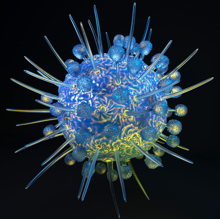
According to the computer simulations developed by the scientists, the virus acts like a Trojan horse, hiding a spike-shaped glycoprotein that it carries on its surface, called hemagglutinin. When hemagglutinin comes into contact with host cells, it releases fusion peptides, which initiate the infection. Hemagglutinin remains completely folded at the start of the process. It was this process that interested the scientists. "It may be the only case known to human beings where a protein starts at a fixed point and literally completely refolds," said Jianpeng Ma, a biochemist at Baylor College of Medicine, in a press release.
Proteins, which build living tissue, are usually made of about 20 amino acids. These amino acids fold and twist to create different sequence combinations. Using the so-called energy landscape theory by co-author Jose Onuchic, the researchers were able to calculate how much energy each amino acid needs within the chain, as well as the influence of the surrounding environment as folding progresses. Using this theory, as well as other algorithms, the researchers were able to determine how an unfolded strand of amino acids collapses into a final, functional protein. Earlier attempts to study hemagglutinin's initial and final structures were unsuccessful due to the speed that the transition occurs. But with the energy landscape theory, the team can predict how a protein will fold no matter how fast it happens.
The team found that during the unfolding and refolding of hemagglutinin, part of the protein "cracks" and releases fusion peptides. "The fusion peptides are the most important part of the molecule," said co-author Jeffrey Noel in the release. "The hemagglutinin is attached to the viral membrane, and when these peptides are released, they embed themselves in the target cell's membrane, creating a connection between the two.”
When this happens, polysaccharide receptors on host cells recognize the hemagglutinin, and the whole virus is absorbed into the cell. Initially, part of the protein forms a cap that protects the segments inside. Acidic conditions in the cell then cause the cap to fall off, and the protein begins to unfold and then refold itself into a completely new shape. "The release of the fusion peptide, which is initially hidden inside hemagglutinin, is triggered by that giant conformational change," Ma said.
Using X-ray crystallography, the team can now watch each step as hemagglutinin goes from being folded to unfolded and back, and the release of peptides during the process.
The virus has proven to be difficult to find a treatment for because it's constantly undergoing mutations in its cap. But the inner core of the protein may be more conserved, said Ma. "We're targeting the part that the virus cannot afford to change," he said. "Therefore, it provides more hope for developing therapeutic agents." Such agents could lead to a universal flu vaccine that would last a lifetime.
Besides influenza, the HIV and Ebola viruses also have a membrane fusion mechanism. Therefore, a comprehensive study of the influenza virus may also lead to understanding treatments for these other viruses.
Source: Lin X, Eddy N, Noel J, et al. Order and disorder control the functional rearrangement of influenza hemagglutinin. PNAS. 2014.


