


What Is Bioengineering? Sometimes It Means Creating A Functional 3D Brain Tissue Model For Studying Disease
Bioengineers are those scientists who use engineering principles to analyze and solve problems in biology and medicine. They are the problem solvers of science, standing squarely at the intersection where biology, chemistry, physiology, and medicine meet. Among the most recent feats in their extraordinary field, a team of bioengineers at Tufts University have managed to create a 3D brain-like tissue.
Not only does the new tissue function just like real brain tissue found in rats, but more importantly, it can be kept alive in a lab for more than two months. Scientists say this new tissue may soon serve as a highly-accurate model for studying normal brain function as well as brain injury and disease, all of which should soon result in new medications for brain dysfunction.
White and Grey
The brain is our most exceptional organ — it is the source of our every passing thought and choreographer of both our sensations and movements. In the brain, grey matter is comprised mostly of neuron cell bodies, while white matter is made up of bundles of axons, which are the projections neurons send out to connect and communicate with one another. Because brain injuries and diseases often affect these areas differently, scientists require models that are capable of showing the grey and white compartmentalization. Until now, they’ve grown neurons in petri dishes; these two dimensional brain cells cannot possibly replicate the complexity of segregated regions of grey and white matter found in a living brain.
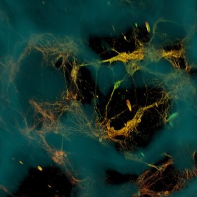
In order to make more realistic brain-like tissue, a team of bioengineers led by Dr. David Kaplan, director of the Tissue Engineering Resource Center at Tufts University, began by creating a unique structure that consisted of two biomaterials with different physical properties: a spongy scaffold made out of silk protein and a softer, collagen-based gel. The scaffold served as a structure onto which neurons could anchor themselves, with the gel encouraging axons to grow through it. Next, the researchers cut the spongy scaffold into a donut shape and populated it with rat neurons to achieve the proper grey-white matter compartmentalization. Then they filled the middle of the donut with the collagen-based gel.
As the team hoped, in just a few days neurons had grown and formed networks around the pores of the scaffold, while sending longer axon projections through the center gel to connect with neurons on the opposite side of the donut. The result was a region of white matter, containing mostly axons, that had formed in the center of the donut. And, separate from this, was a surrounding grey matter, where the cell bodies had gathered.
The first two experiments to demonstrate the tissue’s potential involved studying the chemical and electrical changes that follow traumatic brain injury and tracking changes that occur in response to a drug. Kaplan emphasized the importance of the brain-like tissue's longevity for studying brain disorders. "The fact that we can maintain this tissue for months in the lab means we can start to look at neurological diseases in ways that you can't otherwise," he said. In the meantime, the team is searching for ways to make the tissue model even more brain-like.
Source: Tang-Schomer MD, White JD, Tien LW, et al. Bioengineered functional brain-like cortical tissue. Proceedings of the National Academy of Sciences. 2014.
Published by Medicaldaily.com


