


Baby's Legs Rip Through Mother's Womb After 5 C-Sections Weaken Uterus Wall
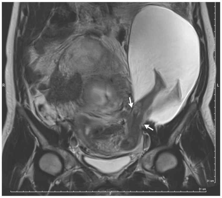
A pregnancy sonogram from France is making headlines for showing the legs of a developing fetus ripping through the mother’s uterine wall. The case is rare — so rare that it’s the first time any of the doctors involved have seen anything quite like it, and only the 26th time it’s been recorded in medical history.
As revealed in a 22nd week ultrasound of a 33-year-old woman, a portion of the amniotic sac measuring 19 by 12 by 9 cm (7.5 by 4.7 by 3.5 inches) had slipped through a hole in the uterine wall. According to the study documenting the case, as a result, the baby's legs up to a little above the knees were allowed to freely kick inside the woman’s body, outside of the protective amniotic sac and uterus walls where they should be.
Read: Is Natural Childbirth Possible After Fibroid Removal Surgery?
By week 30 further sonograms revealed that the tear had increased and even more of the baby’s body was now outside of the uterus. Due to the severity of the situation, and risk to both baby and mother a decision was made to to attempt an early C-section, and at 30 weeks. Thankfully a healthy baby boy weighing 3 lbs was delivered, Science Alert reported.
After the birth, the woman's uterine wall and herniated amniotic sac were treated. At the most recent six months check-up, both the mother and her baby were healthy and doing well.
Doctors believe the mother’s previous history of c-section deliveries may have weakened her uterine wall, increasing the risk of a uterine tear. However this risk usually only presents itself after a woman has had three or four c-section deliveries, Science Alert reported. According to the study, the woman in question had had five previous c-section deliveries.
See Also:
5 Signs You May Be Pregnant Before You Can Take A Test
8 Bizarre Things That Happen While You’re Pregnant For Months Of Surprising Experiences
Published by Medicaldaily.com


