


New Non-Toxic Silver Nanoparticles May Soon Target Cancer Tumors
Silver-based nanoparticles have been used with considerable success for diagnostic and therapeutic purposes in the field of medicine since their invention. But the potential toxicity of their long-term use has always been a concern. The latest nanoparticle, however, invented by a group of scientists at the University of California Santa Barbara, leaves no question about its safety. Not only does it effectively penetrate and target tumor cells — due to its spherical shape and peptide casing — but excess particles that don't hit their target break down and get flushed away, reducing toxicity considerably.
The core of the nanoparticle employs a phenomenon called plasmonics, which reveals its electromagnetic properties. In plasmonics, laser radiation is used to resonate light off of metallic nanoparticles, like gold and silver. This causes so-called "plasmonic enhancements," which concentrate electromagnetic fields on the surface. When dyes are added, they appear far brighter than usual. When the core is etched, the enhancement goes away and the particle becomes dim.
What gives this method an advantage over the others is the etching process. Etching involves selectively removing the excess nanoparticles that do not hit their target. The etching process for this nanoparticle, developed by UCSB's Ruoslahti Research Laboratory, uses biocompatible chemicals to rapidly disassemble and remove the silver nanoparticles outside living cells. This method leaves only the intact nanoparticles for imaging or quantification, thus revealing which cells have been targeted and how much each cell internalized.
"The disassembly is an interesting concept for creating drugs that respond to a certain stimulus," said Gary Braun, a postdoctoral associate in the Ruoslahti Lab at the Department of Molecular, Cellular, and Developmental Biology, in a press release. "It also minimizes the off-target toxicity by breaking down the excess nanoparticles so they can then be cleared through the kidneys."
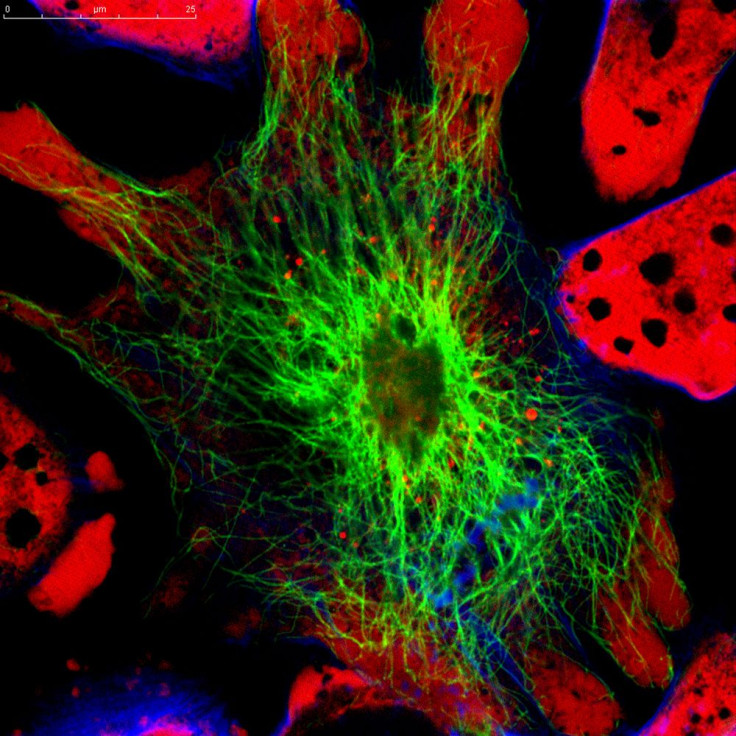
By focusing on the nanoparticles that actually got into cells, we can then understand which cells were targeted and study the tissue transport pathways in more detail."
The intrinsic nature of nanoparticles such as their quantum properties and their ability to absorb and carry other compounds makes them useful in administering drugs that cannot penetrate cell membranes on their own, such as RNA and DNA genetic drugs. These drugs must be taken in through endocytosis, the process by which cells absorb molecules by engulfing them.
"This typically requires a nanoparticle carrier to protect the drug and carry it into the cell," Braun said. "And that's what we measured: the internalization of a carrier via endocytosis."
Since the nanoparticle has a core-shell structure, its exterior peptide coating can be manipulated for effective tumor targeting and internalization. Besides this, silver is known to have several antibacterial properties, even against antibiotic-resistant organisms, and is effective in cancer treatments.
"These new nanoparticles have some remarkable properties that have already proven useful as a tool in our work that relates to targeted drug delivery into tumors," said researcher Erkki Ruoslahti in the release. "They also have potential applications in combating infections. Dangerous infections caused by bacteria that are resistant to all antibiotics are getting more common, and new approaches to deal with this problem are desperately needed. Silver is a locally used antibacterial agent and our targeting technology may make it possible to use silver nanoparticles in treating infections anywhere in the body."
Published by Medicaldaily.com


