


Simple Eye Test For Glaucoma: New Technique Spots Cell Death 10 Years Before Vision Loss Symptoms

We are all at risk for glaucoma; some of us have a higher risk than others. Vision loss is often detected via eye test when permanent damage has already occurred. Now, researchers at the University College London's Institute of Ophthalmology have developed a new technique to detect cell death in the back of the eye, up to a decade before symptoms begin.
Glaucoma develops slowly over several years. During this time, light-sensitive cells of the retinal nerve die due to increased pressure in the eye. This nerve damage leads to the progression of blindness, which is irreversible. The new study published in Brain found a new eye test can detect the earliest signs of glaucoma, which means treatment can start before vision loss begins to deteriorate.
"Although detection has been improving, most patients have lost a third of vision by the time they are diagnosed," said Francesca Cordeiro, lead author of the study, and a professor at UCL Institute of Ophthalmology, in a statement.
He added: "Now, for the first time, we have been able to show individual cell death and detect the earliest signs of glaucoma."
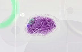
In the clinical trial, Cordeiro and her colleagues recruited eight healthy adults without eye disease and eight adults being treated for early glaucoma in the hospital, without any other eye disease. One of four different doses of fluorescent dye was injected into the bloodstream of patients before researchers scanned their eye with an infrared laser ophthalmoscope. The fluorescent dye sticks to the cells in the retina that are about to die.
This technique is known as DARC, which stands for detection of apoptosing retinal cells. Sick cells show up as fluorescent "white" spots during the eye test up to six hours after injection. Researchers assessed and compared images of those from healthy people and glaucoma patients.
The findings revealed a positive test was linked with a diagnosis of glaucoma and loss of vision later in life. ANX776, the fluorescent marker, was deemed safe and well-tolerated with no side effects. On average, people with glaucoma had more than twice as many white spots that showed dying nerve cells as healthy adults without eye disease. Moreover, people with glaucoma got worse and showed more white spots in the following months compared to those whose disease stayed the same. Those that did not have eye disease tended to have more white spots in old age.
"Loss of sight as you age is an incredibly difficult disability, impacting quality of life and independence," said Bethan Hughes, from Wellcome’s Innovation team, in a statement.
DARC helps detect cell death, and in the early stages of glaucoma, when vision loss is minimal. In addition, this technique means doctors can diagnose patients 10 years earlier than previously believed. Although there's no cure for glaucoma, doctors are able to show individual cell death and spot the earliest signs of the disease.
Researchers hope opticians will eventually do the tests for earlier detection of the disease. This new eye test could be the innovation needed to help people who suffer from vision loss through glaucoma. Currently, the eye test is in Phase I clinical trials — the earliest form of trial meant to check new treatments are safe — but it has only been tested on just 16 people, and far more research is needed, according to the study. The researchers hope to replicate their findings in a larger population for Phase II.
Traditional eye tests may pick up glaucoma, but there's no signs of the disease until patients begin to lose their vision. They can reveal the health of our blood vessels in the eye, and even spot conditions while they're still in their earlier stages, like glaucoma. An optometrist will look for three signs of glaucoma by shining a light into the eye to see the optic disc; test central and peripheral vision; and check the internal pressure with a machine that blows gentle puffs of air into the eye.
In the U.S., the new technique has the potential to help minimize the impact of glaucoma in 3 million patients.
We all need to see it before we believe it.
Source: Cordeiro MF, Normando EM, Cardoso MJ et al. Real-time imaging of single neuronal cell apoptosis in patients with glaucoma. Brain. 2017.
See Also:
Special Contact Lenses Treat Glaucoma, Eliminating Need For Eye Drops
Published by Medicaldaily.com


