


Why Do We Dream? Study Identifies Brain Structure Responsible For Activating REM Sleep And Dreaming
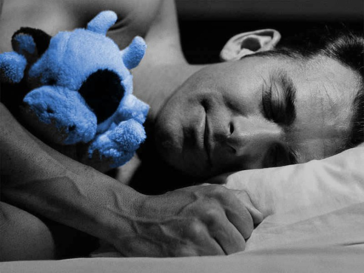
Dreams have been a source of fascination and mystery to both researchers and the general populace alike. There are many things we don’t know about these vivid, mostly involuntary images and ideas that float through our minds while we’re supposed to be resting. The same goes for REM (rapid eye movement) sleep in general — scientists still haven’t been able to pinpoint why this specific part of the sleep cycle is so important, and why our brain waves during REM sleep look so similar to those in a waking state.
Neuroscientists at UC Berkeley, however, have now made a crucial step in discovering how the brain controls REM sleep and dreams. They identified a group of nerve cells that, when activated, immediately trigger a dream state. By activating certain cells located in the medulla, the scientists induced REM sleep in mice within seconds. Inactivating the neurons reduced or even eliminated the animals’ ability to enter REM sleep.
“People used to think that this region of the medulla was only involved in the paralysis of skeletal muscles during REM sleep,” said Yang Dan, a UC Berkeley professor of molecular and cell biology and lead author of the study, in a statement. “What we showed is that these neurons triggered all aspects of REM sleep, including muscle paralysis and the typical cortical activation that makes the brain look more awake than in non-REM sleep.”
This cortical activation is the reason neurons in the waking or REM sleep brain can “fire” more readily than a deeply sleeping brain. Both the brainstem and hypothalamus have been linked to REM sleep, and it’s been suggested that the brainstem is the origin of the electrical and chemical activity that regulates this part of the sleep cycle. The researchers’ results, though, may point to the medulla as the most important area for initiating REM sleep.
“Because of the strong induction of REM sleep — in 94 percent of the recorded trials our mice entered REM sleep within seconds of activating the neurons — we think this might be a crucial node of a relatively small network that makes the decision whether you go into dream sleep or not,” Dan said.
The researchers executed the experiment by inserting a light-sensitive ion channel into specific groups of neurons by way of a virus, a technique called optogenetics. Using a line of genetically engineered mice that had a marker protein in these specific neurons, the team was able to find and stimulate them with precision using a laser light.
These results are not only helpful for understanding the different brain structures responsible for sleeping and dreaming, but they also allow scientists to study why we dream more closely.
“Many psychiatric disorders, especially mood disorders, are correlated with changes in REM sleep, and some widely used drugs affect REM sleep, so it seems to be a sensitive indicator of mental and emotional health,” said first author Franz Weber, a UC Berkeley postdoctoral fellow. “We are hoping that studying the sleep circuit might lead us to new insights into these disorders as well as neurological diseases that affect sleep, like Parkinson’s and Alzheimer’s diseases.”
Dan plans on continuing her studies of the neurons that affect REM sleep, along with those that affect non-REM sleep.
Source: Weber F, Chung S, Beier K, Xu M, Luo L, Dan Y. Control of REM sleep by ventral medulla GABAergic neurons. Nature. 2015.
Published by Medicaldaily.com


